
Polarized Light Microscopy Digital Image Gallery
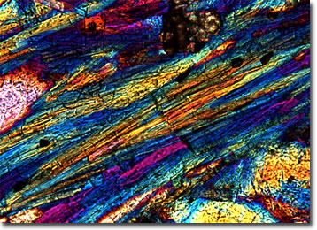
Glaucophane Schist
Glaucophane is a common amphibole, a group of widely distributed rock-forming minerals. The name of the substance stems from the Greek glaukos, meaning “blue,” and fanos, which means "appearing,” a reference to its typical color.
View a second image of Glaucophane Schist
Glaucophane only occurs in certain rocks, such as schist, marble, and eclogite, and is generally formed in a highly metamorphic zone known to geologists by the term blueschist facies. The typical location of blueschist facies is along continental margins affected by shifting of oceanic plates and in areas of high volcanic and seismic activity. Some of the best-known examples of blueschist facies in the world are found in Japan, California, the Mediterranean, and the Alps. These large, beautiful collections of metamorphic rocks gain their moniker from the bluish hue of glaucophane, but also commonly contain jadeite, garnet, muscovite, quartz, lawsonite, and other minerals.
Schist is a type of metamorphic rock that displays a tendency to split into thin layers or flakes. Its name also has Greek roots, evolving from skhistos, meaning “split” or “divisible.” The majority of schists are composed of platy minerals, such as graphite and muscovite, and most exhibit extreme foliation or banding. Schists are particularly abundant among Precambrian rocks and are generally classified according to their mineralogy.