
Polarized Light Microscopy Digital Image Gallery
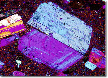
Dacite
Dacite is a form of igneous rock sometimes categorized in its own family and sometimes considered a variety of andesite. Lightly colored, dacite is predominately composed of silica, plagioclase feldspar, and one or more other minerals, such as hornblende, biotite, or augite.
The structure of dacite is most often porphyritic, containing relatively large crystals in a fine-grained matrix called the groundmass. This form of rock may also, however, contain intermediate sized crystals or grains of rounded quartz. Significant deposits of dacite rocks can be found in Spain, Scotland, New Zealand, the Andes mountains, Greece, the western United States, and various other locations throughout the world.
In magma form, dacite is extremely viscous, hindering it from moving very far away from its point of eruption before it cools and solidifies. The substance is, therefore, highly involved in the creation of thick volcanic domes. Dacite, which typically erupts at temperatures that range between 800 and 1000 degrees Celsius, is also one of the rocks most commonly associated with the large volcanic explosions called Plinian eruptions. Named for Pliny the Younger, who in 79 AD recorded the catastrophic effects of the Vesuvius eruption, Plinian eruptions are characterized by the large quantities of solid particles (tephra) and gas they exude high into the stratosphere.