
Polarized Light Microscopy Digital Image Gallery
Biotite Gneiss
Sometimes referred to as black mica, biotite is a common silicate mineral. Similar to other micas, it features a sheetlike structure and exhibits perfect basal cleavage.
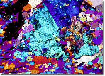
Although a frequent component of metamorphic rocks, such as gneisses, as well as those of igneous origin, biotite is rarely found in sedimentary samples because the mineral has a tendency towards alteration when subjected to chemical weathering. Interestingly, when the mineral is only partially altered, it is sometimes mistaken for gold, its lightened color and flaky appearance being reminiscent of bits of the valuable metallic element. Typically, however, biotite’s dark color prohibits such a mistake.
Gneiss is a medium- to coarse-grained rock that can be found in a variety of locations, but its characteristic banding can perhaps best be seen in massive exposed samples, such as those at Stone Mountain, Georgia and Hickory Nut Gorge in North Carolina. The distinctive alternation of dark and light coloration, which is often an indication of differing proportions of constituent minerals, may be oriented parallel to the ground, but can also occur at an angle. Despite their superficial similarity in appearance to schists, gneisses do not exhibit schistosity, splitting in irregular patterns rather than along planes.