Microscopy Primer
Light and Color
Microscope Basics
Special Techniques
Digital Imaging
Confocal Microscopy
Live-Cell Imaging
Photomicrography
Microscopy Museum
Virtual Microscopy
Fluorescence
Web Resources
License Info
Image Use
Custom Photos
Partners
Site Info
Contact Us
Publications
Home

The Galleries:

Photo Gallery
Silicon Zoo
Pharmaceuticals
Chip Shots
Phytochemicals
DNA Gallery
Microscapes
Vitamins
Amino Acids
Birthstones
Religion Collection
Pesticides
BeerShots
Cocktail Collection
Screen Savers
Win Wallpaper
Mac Wallpaper
Movie Gallery

Phase Contrast Image Gallery

Tobacco Blue Mold

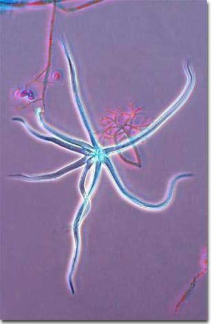
Blue mold affects only tobacco plants and is a local, regional, and continental problem. It is caused by the fungus Peronospora tabacina, which rapidly produces tiny spores that are ejected into the air and spread by air currents. As evidenced by this micrograph, combining phase contrast microscopy with classical histological staining techniques often yields enhancement of cellular features.

In recent years, biologists have begun using meteorological tools to track clouds of these spores traveling on the wind over the Caribbean, Latin America, the continental United States, and Canada. This information is used to predict outbreaks of blue mold, giving tobacco growers an opportunity to protect and treat their crops.

The blue mold fungus produces spores that are the result of both sexual and vegetative reproduction. Resting spores, or oospores, are produced through sexual reproduction (fusion of female and male gametes) and are believed to be more involved with long term survival of the species. Sporangiospores are produced vegetatively (i.e., the fungus makes clones of itself) and can be made by the millions within hours after a tobacco plant is infected.

BACK TO THE PHASE CONTRAST GALLERY

Questions or comments? Send us an email.
© 1998-2022 by Michael W. Davidson and The Florida State University. All Rights Reserved. No images, graphics, scripts, or applets may be reproduced or used in any manner without permission from the copyright holders. Use of this website means you agree to all of the Legal Terms and Conditions set forth by the owners.
This website is maintained by our
Graphics & Web Programming Team
in collaboration with Optical Microscopy at the
National High Magnetic Field Laboratory.
Last modification: Friday, Nov 13, 2015 at 02:19 PM
Access Count Since March 15, 2000: 28840
For more information on microscope manufacturers,
use the buttons below to navigate to their websites: