
Phase Contrast Image Gallery
Stomach Cancer
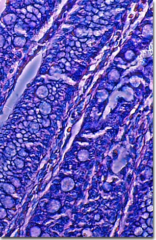
A stained thin section of human stomach reveals damage caused by carcinoma. As evidenced by this micrograph, combining phase contrast microscopy with classical histological staining techniques often yields enhancement of cellular features.
Until after World War II, stomach cancer, also known as gastric cancer or gastric carcinoma, was one of the leading cancer killers in the United States. While it's much less prevalent in the US now, it is still one of the most common and lethal cancers in Japan, Korea, Latin America, and Eastern Europe.
Stomach cancer is primarily an acquired form of cancer, affecting mostly people in their 60s and 70s. A higher incidence is associated with diets containing large amounts of smoked foods, salted fish and meat, certain foods high in starch that are also low in fiber, and pickled vegetables. Eating whole grain products, fresh fruits and vegetables that contain vitamins A and C appears to lower the risk of stomach cancer. Other environmental factors such as tobacco and alcohol use, previous stomach surgery, and infection with Helicobacter pylori bacteria, are also associated with an increased incidence.
Some people may have a hereditary predisposition to this form of cancer, however. People who are blood type A have a higher risk of developing stomach cancer, as do people with a gene mutation that can cause Lynch Syndrome, or hereditary nonpolyposis colon cancer.
BACK TO THE PHASE CONTRAST GALLERY