
Phase Contrast Image Gallery
Squamous Cell Papilloma
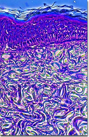
A stained thin section of human elpithelial tissue exhibiting damage from squamous cell papilloma virus is illustrated below. As evidenced by the micrograph, combining phase contrast microscopy with classical histological staining techniques in pathological research often yields enhancement of cellular features.
Papillomas, also known as warts or corns, are benign (noncancerous) growths of the skin or mucous membranes and are caused by the Human Papilloma Virus (HPV). HPVs are very common and are only able to infect the epithelium of the skin.
While innocuous for the most part, some strains of this virus are associated with cellular changes that can eventually lead to cancer. There are two kinds of abnormal tissues caused by HPV that physicians can identify: condyloma (warts) and dysplasia (pre-cancer). While there is no cure for the virus at present, it generally does not require treatment. Most papillomas will go away on their own or can be tolerated if they are not in a pre-cancerous state. Surgical removal is the only treatment for disfiguring or potentially cancerous papillomas.
Over 65 virus types have been identified so far, based on DNA analysis. The genome of the virus is contained entirely on a double-stranded circular DNA molecule, which replicates entirely in the nucleus of the host cell. Usually, it replicates independently of the host genome, but will integrate into the DNA of the host cell on occasion, which could eventually make it a candidate as a vehicle for gene therapies.
BACK TO THE PHASE CONTRAST GALLERY