Microscopy Primer
Light and Color
Microscope Basics
Special Techniques
Digital Imaging
Confocal Microscopy
Live-Cell Imaging
Photomicrography
Microscopy Museum
Virtual Microscopy
Fluorescence
Web Resources
License Info
Image Use
Custom Photos
Partners
Site Info
Contact Us
Publications
Home

The Galleries:

Photo Gallery
Silicon Zoo
Pharmaceuticals
Chip Shots
Phytochemicals
DNA Gallery
Microscapes
Vitamins
Amino Acids
Birthstones
Religion Collection
Pesticides
BeerShots
Cocktail Collection
Screen Savers
Win Wallpaper
Mac Wallpaper
Movie Gallery

Phase Contrast Image Gallery

Snake Skin

A stained thin section of snake skin imaged using phase contrast optics is illustrated below. The image was captured using an Olympus IX-70 inverted microscope equipped with a DP-10 digital camera.

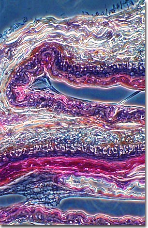
Contrary to popular belief snake skin is dry, not slimy (with the exception of newly born or hatched snakes). Snake skin consists entirely of overlapping scales, specialized folds of skin, that provide protection from the environment and from predators. The scales are composed primarily of keratin, but also contain waxes that help to prevent water loss through the skin.

Epidermis is not replaced on an ongoing basis in snakes as it is with other animals. When the snake grows too large for its skin, or the skin begins to wear out, it is shed in one piece. This molting process can happen several times a year, if a snake is young and growing, or it may just occur once a year to replace worn out skin.

The pattern, type and configurations of scales of shed snake skins are as unique and distinctive as finger prints are amongst humans. People with sufficient expertise can usually identify the species of snake, or the group it belongs to, simply by examining a molted skin.

BACK TO THE PHASE CONTRAST GALLERY

Questions or comments? Send us an email.
© 1998-2022 by Michael W. Davidson and The Florida State University. All Rights Reserved. No images, graphics, scripts, or applets may be reproduced or used in any manner without permission from the copyright holders. Use of this website means you agree to all of the Legal Terms and Conditions set forth by the owners.
This website is maintained by our
Graphics & Web Programming Team
in collaboration with Optical Microscopy at the
National High Magnetic Field Laboratory.
Last modification: Friday, Nov 13, 2015 at 02:19 PM
Access Count Since April 15, 2000: 45036
For more information on microscope manufacturers,
use the buttons below to navigate to their websites: