
Phase Contrast Image Gallery
Sebaceous Cysts
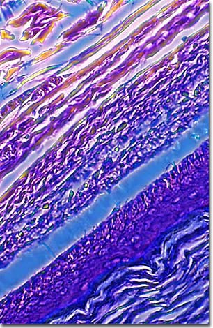
Illustrated below is a phase contrast image of a human sebaceous cyst thin section stained with eosin and hematoxylin. A sebaceous cyst occurs when keratin-producing glands in the skin become clogged, causing the gland to enlarge.
Also referred to as a wen, epidermal cyst, or pilar cyst, it is caused by plugged ducts in malformed hair follicles and generally requires no medical treatment. Occasionally, one may become enlarged, infected, or persistent enough to warrant surgical removal. Sebaceous cysts grow slowly and only rarely become malignant (cancerous).
The term "sebaceous" cyst is something of a misnomer because these cysts do not, in fact, occur in the sebaceous glands but in the skin's hair (keratin) producing follicles. Dermatologists now also use the term trichilemnal cysts to describe them. Keratin is the fibrous structural protein that, in humans, makes up hair, nails, and the outermost layers of skin. In sebaceous cysts, it is a cheesy-looking, semisolid material.
Sebaceous glands produce sebum, an oily substance of unknown function though it undoubtedly has significance since humans have more and larger sebum-producing sebaceous glands than most mammals. These glands are the ones that produce whiteheads and blackheads, which are not sebaceous cysts.
BACK TO THE PHASE CONTRAST GALLERY