
Phase Contrast Image Gallery
Multipolar Neurons
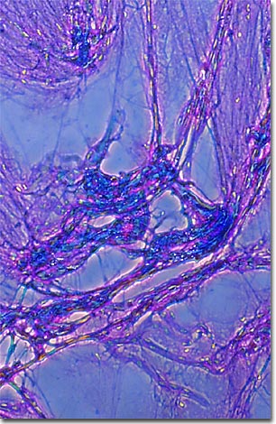
A cytological smear of human multipolar neurons stained with a mixture of eosin and hematoxylin is illustrated in the photomicrograph presented below. As evidenced by the micrograph, combining phase contrast microscopy with classical histological staining techniques in pathological research often yields enhancement of cellular features.
The human nervous system is structurally and functionally divided into the peripheral nervous system, a conglomerate of neurons and nerve tissues that transmit signals back and forth between peripheral organs and the central nervous system, which includes the brain and spinal cord. Information is processed in the brain and integrated with existing information to form an appropriate response to external stimuli.
A typical human brain contains over 100 billion neurons, about 10 percent of the total number of cells in the entire nervous system. Neurons, the functional component of the nervous system, are elongated cells that exhibit a high degree of conductivity, which allows them to sense environmental changes and transmit chemical impulses from one part of the body to another.
Neurons consist of a cell body and one or more cytoplasmic processes that extend from the cell body. The two main types of neuron processes are dendrites, which detect environmental changes from the eyes, ears, nose, and tactile stimuli, and the axons, which conduct electrochemical impulses. Neuron processes are used to classify individual cell types. Those neurons having unbranched dendrites are known as monopolar, whereas those having branched axons are termed multipolar neurons.
BACK TO THE PHASE CONTRAST GALLERY