
Phase Contrast Image Gallery
Fish Gills
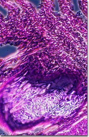
Fish gotta swim but they gotta breathe too. Unlike land vertebrates or marine mammals, they don't have lungs, but they do have paired respiratory structures called gills, also called branchia. The photomicrograph below illustrates a stained thin section of fish gill imaged using phase contrast optics.
Outgrowths of the body wall, gills remove dissolved oxygen from water and expel carbon dioxide waste from the bloodstream. This is how fish can breathe underwater without ever having to come to the surface for air. When there are insufficient quantities of dissolved oxygen in the water, they will suffocate.
Not all fish species rely entirely on their gills. Some, especially when they are young, absorb a large proportion of the oxygen they need through their skin. In a few other species, the air bladder, which most modern fish use as a ballast organ to control their depth, is specialized as an accessory breathing organ or lung. These fish are obligate air breathers and will drown if they cannot breathe air, even in well-oxygenated water. The air bladder, not gills, is the structure that gave rise to the evolution of the lung and follows the same developmental pattern as the lungs of land vertebrates.
Fish are the most diverse vertebrate group and make up about half of all known vertebrate species. More fish species are being discovered at a rate of 200 to 300 a year.
VIEW THIS SPECIMEN UNDER FLUORESCENCE ILLUMINATION
BACK TO THE PHASE CONTRAST GALLERY