
Phase Contrast Image Gallery
Diabetes Mellitus
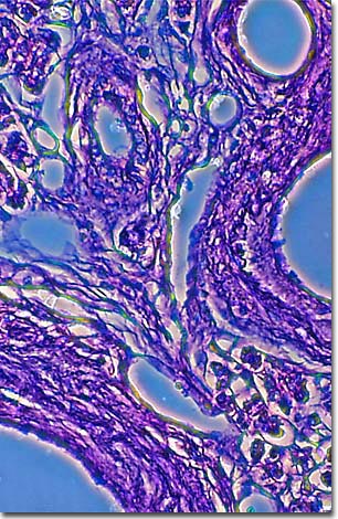
A stained thin section of human pancreas reveals damage caused by the devistating disease diabetes mellitus. As evidenced by this micrograph, combining phase contrast microscopy with classical histological staining techniques often yields enhancement of cellular features.
Known since antiquity, diabetes mellitus is widely recognized as one of the leading causes of death and disability in the United States. Also referred to as sugar or sugar diabetes, it is an incurable, and potentially fatal, metabolic disorder that creates an energy crisis in the body.
Most of the food that a person eats is digested and broken down into a simple sugar called glucose. Glucose molecules are released into the bloodstream during digestion and, with the help of the hormone insulin, are transported into individual body cells to use as fuel. Diabetes occurs when the body is unable to produce insulin or utilize it. The body's cells become starved for glucose, even though ample quantities are available in the bloodstream. There are three types of diabetes, Type I insulin-dependent, Type II noninsulin-dependent, and gestational diabetes.
Insulin dependents, about 5-10% of all diabetics, have lost the ability to produce insulin and require insulin injections for their survival. Until the 1920s, when insulin was isolated, Type I diabetics typically died within a short time after onset of the disease.
Generally, Type I diabetes comes on suddenly and is seen initially in people under the age of 30. It is caused by damage to the pancreas, a hand-sized organ near the stomach that helps with digestion and produces insulin. Areas of the pancreas, called the islets of Langerhans, lose their ability to produce insulin because their insulin-producing beta cells have been destroyed. While it is not yet known what is killing the beta cells, there may be a variety of causes, such as genetic predispositions, autoimmune problems or viruses.
Type II diabetes affects 90-95% of all diabetics, comes on gradually, and is associated with obesity and aging. Type II diabetics produce insulin but their body cells have lost the ability to respond to insulin. The end result is the same as it is for Type I diabetics--too much sugar in the blood. Type II can be controlled with diet and exercise, but insulin injections may be required in severe cases.
Gestational diabetes is the only form of diabetes that is temporary. For reasons not yet understood, it develops during pregnancy and usually disappears after the childbirth. Even if they fully recover, however, women with this form are generally inclined to develop Type II diabetes later in life.
BACK TO THE PHASE CONTRAST GALLERY