Microscopy Primer
Light and Color
Microscope Basics
Special Techniques
Digital Imaging
Confocal Microscopy
Live-Cell Imaging
Photomicrography
Microscopy Museum
Virtual Microscopy
Fluorescence
Web Resources
License Info
Image Use
Custom Photos
Partners
Site Info
Contact Us
Publications
Home

The Galleries:

Photo Gallery
Silicon Zoo
Pharmaceuticals
Chip Shots
Phytochemicals
DNA Gallery
Microscapes
Vitamins
Amino Acids
Birthstones
Religion Collection
Pesticides
BeerShots
Cocktail Collection
Screen Savers
Win Wallpaper
Mac Wallpaper
Movie Gallery

Phase Contrast Image Gallery

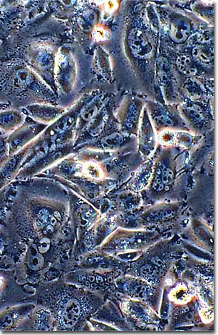
Chinese Hamster Ovary Cells

This monolayer tissue culture of Chinese hamster ovarian fibroblast cells was photographed in a plastic culture vessel with phase contrast optics using an Olympus inverted microscope.

Most animal cells display a finite lifetime when isolated and grown in a tissue culture medium that supplies necessary nutrients, salts, and vitamins. Typical vertebrate cells divide between 50 and 100 times before they fail to continue cell division and eventually die. Many theories suggest that this limited life span is related to the corresponding life cycle of the parent organism from which the cultured cells were derived.

Occasional changes in the genetic makeup of cultured cells allow them to propogate indefinitely, making them effectively immortal. Such lines are said to be transformed, and are often used in research as a standardized cell line. Chinese hamster ovary (CHO) cells were introduced in the early 1960s as a viable epithelial cell line containing twin female X chromosomes. The most common variety of CHO cells has a nutritional requirement for the amino acid proline, which makes this cell line an ideal candidate for genetic studies.

BACK TO THE PHASE CONTRAST GALLERY

Questions or comments? Send us an email.
© 1998-2022 by Michael W. Davidson and The Florida State University. All Rights Reserved. No images, graphics, scripts, or applets may be reproduced or used in any manner without permission from the copyright holders. Use of this website means you agree to all of the Legal Terms and Conditions set forth by the owners.
This website is maintained by our
Graphics & Web Programming Team
in collaboration with Optical Microscopy at the
National High Magnetic Field Laboratory.
Last modification: Friday, Nov 13, 2015 at 02:19 PM
Access Count Since October 16, 1999: 40861
For more information on microscope manufacturers,
use the buttons below to navigate to their websites: