Fluorescence Digital Image Gallery
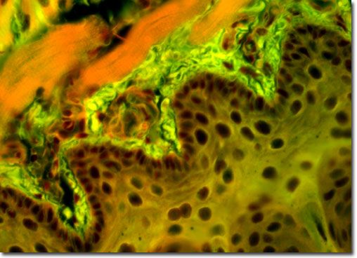
Trichina Worm
|
The trichina worm lives inside the small intestine of a host animal, where it mates and reproduces. Once her eggs have been fertilized, the female burrows into the intestinal wall and releases her larvae. The larvae migrate into the lymph channels of the intestine, from which they enter the bloodstream and travel to all parts of the body. When the larvae reach the skeletal muscles they burrow into the muscles and form tough cyst-like cocoons. The host secretes lime salts, which are deposited in the capsule, eventually transforming the capsule into a completely calcified cyst. The worms may live in the cyst for years until they are consumed and digested by another mammal. |
© 1995-2022 by Michael W. Davidson and The Florida State University. All Rights Reserved. No images, graphics, software, scripts, or applets may be reproduced or used in any manner without permission from the copyright holders. Use of this website means you agree to all of the Legal Terms and Conditions set forth by the owners.
This website is maintained by our
|