
Fluorescence Digital Image Gallery
Snail Radula
Snails belong to the class Gastropoda, the largest group of the mollusk phylum. The most recent estimate of the number of known gastropod species is 37,500. Although gastropods are common in marine and freshwater habitats, they are the only mollusks to flourish on land.
Snails have a large foot for creeping along surfaces, a single coiled shell that encloses the organs, and a head with eyes and tentacles. Like most other gastropods, snails feed by using a specialized rasping organ called a radula. It is a ribbon-like structure covered with small horny teeth called denticles that tear food into pieces that are then collected by lips or a proboscis. New denticles are constantly being produced to replace those worn away at the front. Snails may be herbivorous or carnivorous, predatory or parasitic. Some feed on plankton and deposits of detritus filtered from the water.
The gastropods are a highly successful group of animals and exhibit great diversity in size and structure. The smallest species are barely visible, while the California black sea hare can weigh as much as 30 pounds. Most have a shell, but sea slugs and garden slugs have lost their shells over the eons. One species of snail has a shell with two halves, like that of a bivalve.
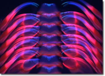
The specimen presented here was imaged with a Nikon Eclipse E600 microscope operating with fluorite and/or apochromatic objectives and vertical illuminator equipped with a mercury arc lamp. Specimens were illuminated through Nikon dichromatic filter blocks containing interference filters and a dichroic mirror and imaged with standard epi-fluorescence techniques. The filter combination utilized to image the snail radula stained thin section was a UV-2E/C and Texas Red HYQ. Photomicrographs were captured with an Optronics MagnaFire digital camera system coupled to the microscope with a lens-free C-mount adapter.
BACK TO THE FLUORESCENCE DIGITAL IMAGE GALLERY