
Fluorescence Digital Image Gallery
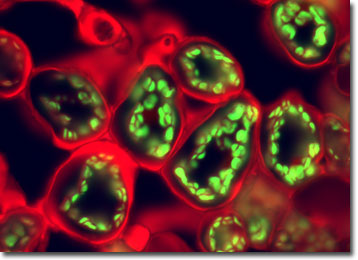
Privet Leaf
About 50 species of privet shrubs and small trees belong to the genus Ligustrum of the olive family, Oleaceae. They are popularly used as hedges and screens. Privets are native to Europe, Asia, and the Mediterranean regions.
The common privet (L. vulgare) is native to northeastern Europe and Great Britain, but has become naturalized in areas of North America after years of being used as an ornamental plant. Privet grows quickly, reaching heights of 15 feet, and is exceptionally resilient. Its flowers bloom in clusters, or panicles, of small white blossoms.
Small-leaf privet (L. sinense) and broadleaf privet (L. lucidum) were introduced to Australia from Asia, and are now causing widespread problems as an invasive species on the New South Wales north and central coast. They typically infest disturbed (human modified) habitats, as well as native bush land and gardens. Many areas in New South Wales receive more water today, through irrigation for farming and urban land use, than naturally occurred before Europeans settled the region. Privet root systems have a remarkable ability to absorb nutrients and moisture from the soil, which makes it difficult to replant other species in the area after extracting the privets. Also, the pollen produced from this species is causing allergic reactions in many people.
The specimen presented here was imaged with a Nikon Eclipse E600 microscope operating with fluorite and/or apochromatic objectives and vertical illuminator equipped with a mercury arc lamp. Specimens were illuminated through Nikon dichromatic filter blocks containing interference filters and a dichroic mirror and imaged with standard epi-fluorescence techniques. Specific filters for the privet leaf stained thin section were a B-2E/C and a Y-2E/C. Photomicrographs were captured with an Optronics MagnaFire digital camera system coupled to the microscope with a lens-free C-mount adapter.
BACK TO THE FLUORESCENCE DIGITAL IMAGE GALLERY