
Fluorescence Digital Image Gallery
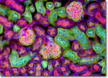
Mouse Kidney Thin Section
The kidney is an organ that maintains water balance and expels metabolic wastes in vertebrates and some invertebrates. Primitive and embryonic kidneys have sets of specialized tubules that empty into two collecting ducts that pass urine into a primitive bladder. The more advanced mammalian kidney is a paired compact organ with functional units, called nephrons, that filter the blood, reabsorbing water and nutrients and secreting wastes, producing the final urine.
Mouse kidneys are located on the dorsal (upper) wall of the abdominal cavity and are securely held in place by fibrous capsules. Like other mammalian kidneys, the outer layer of the kidney is brownish red and granular in appearance, with a firm consistency. Mouse kidneys are similar to human kidneys, which is why they are often used to simulate human kidneys in scientific studies. Mice have played a significant role in experiments used to diagnose the possible cause and treatment of IgA nephropathy, or Berger's ("burrjays") disease. This is the most common non-diabetic kidney disease and affects as many as two to four percent of the world's population.
Laboratory mice are special breeds of house mice and are used in many scientific experiments because of their close mammalian relationship to humans. Compared to larger mammals, mice and other rodents are small, easy to handle, inexpensive to house, and breed quickly. During the twentieth century, scientists bred different strains of mice with genetic deficiencies in order to produce models for human diseases.
The specimen presented here was imaged with a Nikon Eclipse E600 microscope operating with fluorite and/or apochromatic objectives and vertical illuminator equipped with a mercury arc lamp. Specimens were illuminated through Nikon dichromatic filter blocks containing interference filters and a dichroic mirror and imaged with standard epi-fluorescence techniques. Specific filters for the mouse kidney stained thin section were a UV-2E/C, B-2E/C, and a Y-2E/C. Photomicrographs were captured with an Optronics MagnaFire digital camera system coupled to the microscope with a lens-free C-mount adapter.
BACK TO THE FLUORESCENCE DIGITAL IMAGE GALLERY