
Fluorescence Digital Image Gallery
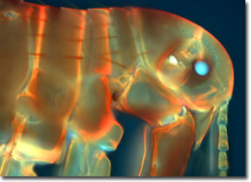
Human Flea
Pulex irritans, the human flea, is one of more than 1,600 species and subspecies of fleas that populate the Earth from the Arctic Circle to the deserts of Africa. Fleas belong to the insect order Siphonaptera and parasitize mammals and birds for their blood, using specialized anatomical structures to attach to the hosts' skin.
As its common name suggests, the preferred food for Pulex is human blood, but it will feed on other mammals as well. Historically, human populations have had ongoing infestations of Pulex, and the problem has yet to completely come to an end. Infestations of Pulex still occur in some areas, but they are less frequent in societies where personal hygiene dictates frequent bathing and clothes laundering.
The human flea is not the primary species responsible for transmitting the bubonic plague throughout Europe during the Middle Ages (that dubious honor belongs to the rodent flea), although it is capable of spreading it. One disease that Pulex is known to transmit, however, is murine typhus, a mild form of typhus caused by the bacterium Rickettsia prowazekii. The flea becomes infected by feeding on a human who has the disease and the bacteria grows in the epithelial cells lining the flea's gut wall, eventually being excreted in the insect's feces. The infection kills the flea after 12 to 18 days. A person becomes infected by scratching the flea bite, thus rubbing the flea's infected feces into the wound. After an incubation period of one to two weeks, an infected person experiences headaches, loss of appetite, malaise, and a rapid rise in temperature with fever, chills, marked prostration, and nausea. Four to six days later a rash appears over most of the body.
The flea's life cycle consists of four stages: egg, larva, pupa, and adult. Eggs are laid on the body of the host animal, but since they are not sticky, they can drop in any number of places. The larva resembles a small legless caterpillar and it feeds on dried excrement, dried bits of skin, dead mites, dried blood, and other organic debris. Fecal matter from the parent flea is essential to the successful metamorphosis of some species of flea larvae. During this time the parent flea consumes a great deal of blood, up to 30 times its own weight, to produce a large quantity of feces for its larvae. After three molts, the larvae spin a silk cocoon that begins the pupal stage. The pupae emerge as adults days, weeks, or even months later. If conditions are unfavorable, a cocooned flea can remain dormant for up to a year, waiting for warm-blooded creatures to prey upon. At any given time, only five percent of living fleas are in adult form, while most are in the egg, larva, or pupa stages. The life span of adult fleas varies from several weeks to over a year.
The specimen presented here was imaged with a Nikon Eclipse E600 microscope operating with fluorite and/or apochromatic objectives and vertical illuminator equipped with a mercury arc lamp. Specimens were illuminated through Nikon dichromatic filter blocks containing interference filters and a dichroic mirror and imaged with standard epi-fluorescence techniques. Specific filters for the human flea specimen were a UV-2E/C, B-2E/C, and a Y-2E/C. Photomicrographs were captured with an Optronics MagnaFire digital camera system coupled to the microscope with a lens-free C-mount adapter.
BACK TO THE FLUORESCENCE DIGITAL IMAGE GALLERY