
Fluorescence Digital Image Gallery
Head Lice
There are three species of louse that parasitize humans: the head louse (Pediculus humanus capitis); the body louse, better known as the cootie (P. humanus humanus); and the crab, or pubic, louse (Phthirus pubis). These species belong to the insect order Anoplura and have long been associated with people. Archaeologists have even found louse combs buried with Egyptian mummies, who were apparently anticipating head lice in the afterlife.
The head louse feeds on human blood several times daily and resides close to the scalp to maintain its body temperature. Lice can be transmitted by contaminated items, such as hats, combs, sports equipment, and pillowcases. They may also remain on bedding or upholstered furniture for a brief period, although they will live for only 48 hours once they fall off the host's head.
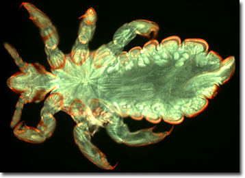
Head lice occur in three stages: the nit, the nymph, and the adult. Nits are small, oval-shaped, yellowish eggs. Often they are difficult to see and can be confused with dandruff or droplets of hair spray. After one week the nits hatch into nymphs, baby lice. Nymphs look like small adult lice, but are only about the size of a pinhead. They mature after seven to 14 days of feeding on blood. About the size of a sesame seed, the adult louse can be seen with the naked eye. They live about 30 days, securing themselves to individual hairs with claws found at the end of each of their six legs. They can also be found behind the ears and near the neckline at the back of the neck. An adult female louse may deposit more than 100 eggs in her lifetime at a rate of about six eggs each day.
Head lice infections are very common; six to 12 million people worldwide are infected annually. A tickling sensation, itching, excessive scratching, and sores can be signs of an infestation, but a medical diagnosis is often necessary to rule out other causes. Luckily, head lice are treatable, although many strains are becoming resistant to the current drugs of choice.
Head lice have also embellished the English language (and other languages, undoubtedly) as a result of their long and close association with humans. Calling someone a "nitwit" is the same as saying he has the intelligence of a louse egg. A person may be considered "lousy," but the word no longer means that she's infested with lice. Phrases, such as "getting down to the nitty gritty" or "nit-picking," are age-old references to the detailed business of removing louse eggs or nits.
The specimen presented here was imaged with a Nikon Eclipse E600 microscope operating with fluorite and/or apochromatic objectives and vertical illuminator equipped with a mercury arc lamp. Specimens were illuminated through Nikon dichromatic filter blocks containing interference filters and a dichroic mirror and imaged with standard epi-fluorescence techniques. Specific filters for the head louse specimen were a UV-2E/C, B-2E/C, and a Y-2E/C. Photomicrographs were captured with an Optronics MagnaFire digital camera system coupled to the microscope with a lens-free C-mount adapter.
BACK TO THE FLUORESCENCE DIGITAL IMAGE GALLERY