
Fluorescence Digital Image Gallery
Elderberry (Sambucus canadensis)
About 30 species of elderberry make up the genus Sambucus of the family Caprifoliaceae. Elderberry bushes can be found in most forested temperate or subtropical areas around the world. In horticulture, the bushes are often used as garden shrubs and are well known for their fruit, which is used to make wines, syrups, cordials, jellies, pies, and also serves as a source of food for wildlife.
A number of varieties of the American elderberry (Sambucus canadensis), which grows up to eight feet tall, have been cultivated for home or commercial use. The berries of the plant arrive after the clusters of white flowers and feature an array of colors including red, blue, black and yellow. The unopened flower buds can be pickled and used as a substitute for capers. Traditionally, the berries were used as a source of various violet dyes. Black dye was also made from the bark and green dye from the leaves. Additionally, all parts of the plant were, throughout various times and cultures, believed to have healing properties. Bark and root extracts were used as laxatives, leaf extracts were used as ointments for bruises, and flower water was used in eye and skin lotions. In modern medicine, the flowers are used to reduce fevers and used as a diuretic and expectorant.
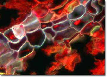
The photomicrograph presented above displays a lenticel of an elderberry plant. A lenticel is a loose aggregation of cells that penetrate the surface of a woody plant. Upon close inspection, it appears to be a blister-like break in the surface. These cells exchange gases between the atmosphere and the underlying tissues. Each lenticel becomes a pathway through which oxygen and other gases can diffuse to the living cells of the bark. Without sufficient oxygen, the cells of bark would die.
The specimen presented here was imaged with a Nikon Eclipse E600 microscope operating with fluorite and/or apochromatic objectives and vertical illuminator equipped with a mercury arc lamp. Specimens were illuminated through Nikon dichromatic filter blocks containing interference filters and a dichroic mirror and imaged with standard epi-fluorescence techniques. Specific filters for the elderberry lenticel specimen were a UV-2E/C, B-2E/C, and a Y-2E/C. Photomicrographs were captured with an Optronics MagnaFire digital camera system coupled to the microscope with a lens-free C-mount adapter.
BACK TO THE FLUORESCENCE DIGITAL IMAGE GALLERY