Fluorescence Digital Image Gallery
Normal African Green Monkey Kidney Epithelial Cells (Vero)
|
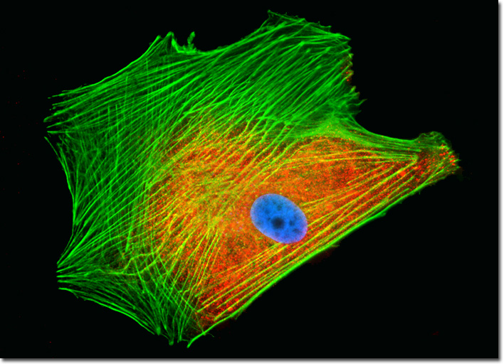
Cytoskeletal actin filaments are typically nucleated at the plasma membrane, rather than the centrosome that acts as an organizational center for microtubules. Thus, it is the cell periphery that generally contains the highest concentration of actin filaments. When located directly underneath the plasma membrane, actin filaments comprise part of the cell cortex, which regulates the form and motion of the surface of the cell, playing a key role in whether or not a cell develops projections such as filopodia or microvilli. A number of external factors generally control actin filament nucleation and enable the cytoskeletal filaments to change their characteristics rapidly upon signaling. A group of special proteins, two of which are actin-related proteins (ARPs) that are very similar to actin, are chiefly responsible for nucleation catalysis. This ARP complex is most prominent in locations where actin filament growth needs to be a brisk process, and is associated with various signaling molecules and components of the plasma membrane. The log phase culture of Vero cells illustrated above was fixed, permeabilized, blocked, and treated with mouse anti-C-protein (myocardial) monoclonal primary antibodies followed by goat anti-mouse secondary antibodies conjugated to Alexa Fluor 568. Mixed in with the secondary reagent was Alexa Fluor 488 conjugated to phalloidin to label the filamentous actin cytoskeleton. Nuclei were counterstained with Hoechst 33258. Images were recorded in grayscale with a QImaging Retiga Fast-EXi camera system coupled to an Olympus BX-51 microscope equipped with bandpass emission fluorescence filter optical blocks provided by Omega Optical. During the processing stage, individual image channels were pseudocolored with RGB values corresponding to each of the fluorophore emission spectral profiles. |
© 1995-2022 by Michael W. Davidson and The Florida State University. All Rights Reserved. No images, graphics, software, scripts, or applets may be reproduced or used in any manner without permission from the copyright holders. Use of this website means you agree to all of the Legal Terms and Conditions set forth by the owners.
This website is maintained by our
|