Fluorescence Digital Image Gallery
Normal African Green Monkey Kidney Epithelial Cells (Vero)
|
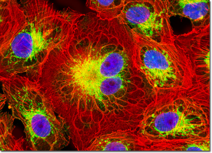
Lectins were discovered in plants over a century ago, and are known for their ability to clot or agglutinate (aggregate) red blood cells, as well as many subcellular organelles, because of the glycoprotein-rich membranes. Isolated from Glycine max (the common soybean) seeds, soybean agglutinin (SBA) is a lectin protein composed of four approximately equal subunits. The tetrameric protein is approximately 120,000 Daltons in weight and has saccharide binding sites that recognize and bind terminal alpha- and beta-N-acetylgalactosamine and galactopyranosyl oligosaccharides. Conjugates of the lectin SBA are commercially available and serve as versatile probes for detection of cell surface and intracellular glycoproteins in fluorescence microscopy, flow cytometry, and gel electrophoresis. Presented in the digital image above is an adherent culture of Vero cells that was labeled with Oregon Green 488 conjugated to soybean agglutinin, a lectin isolated from Glycine max that selectively binds terminal alpha- and beta-N-acetylgalactosamine and galactopyranosyl residues. In addition, the cells were labeled with Alexa Fluor 568 conjugated to phalloidin and DAPI, targeting the cytoskeletal F-actin network and DNA, respectively. Images were recorded in grayscale with a QImaging Retiga Fast-EXi camera system coupled to an Olympus BX-51 microscope equipped with bandpass emission fluorescence filter optical blocks provided by Omega Optical. During the processing stage, individual image channels were pseudocolored with RGB values corresponding to each of the fluorophore emission spectral profiles. |
© 1995-2022 by Michael W. Davidson and The Florida State University. All Rights Reserved. No images, graphics, software, scripts, or applets may be reproduced or used in any manner without permission from the copyright holders. Use of this website means you agree to all of the Legal Terms and Conditions set forth by the owners.
This website is maintained by our
|