Fluorescence Digital Image Gallery
Human Bone Osteosarcoma Cells (U-2 OS)
|
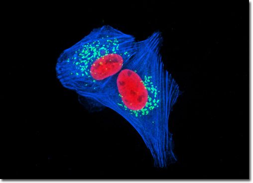
Giantin is a membrane-bound protein resident in the Golgi complex of mammalian cells. The glycoprotein, which can be found in the cis and medial Golgi regions, contains an elongated rod-like cytoplasmic domain that is at least 350 kiloDaltons in size. Highly conserved, giantin may be involved in intercisternal cross-bridge structures that are suspected to play a role in Golgi cisternae stacking. Moreover, studies indicate that endogenous giantin is very static, remaining in pericentriolar Golgi, rather than accumulating on Golgi-derived tubules with Brefeldin A treatment or onto newly formed Golgi mini-stacks upon depolymerization of microtubules. It has been suggested, therefore, that the association of giantin with the Golgi complex is very stable, and that the protein does not recycle to the endoplasmic reticulum in an efficient manner. The U-2 OS cell culture featured above was fixed, permeabilized, washed, and blocked with 10-percent normal goat serum in phosphate-buffered saline prior to immunofluorescent labeling with rabbit primary antibodies to giantin, a protein resident in the Golgi complex of mammalian cells. The culture was subsequently stained with a mixture of secondary antibodies conjugated to Alexa Fluor 488. In addition, histones were immunofluorescently labeled with primary anti-histone mouse monoclonal antibodies followed by goat anti-mouse Fab heavy and light chain fragments conjugated to Texas Red. The filamentous actin network was counterstained with Alexa Fluor 350 conjugated to phalloidin. Images were recorded in grayscale with a QImaging Retiga Fast-EXi camera system coupled to an Olympus BX-51 microscope equipped with bandpass emission fluorescence filter optical blocks provided by Omega Optical. During the processing stage, individual image channels were pseudocolored with RGB values corresponding to each of the fluorophore emission spectral profiles. |
© 1995-2022 by Michael W. Davidson and The Florida State University. All Rights Reserved. No images, graphics, software, scripts, or applets may be reproduced or used in any manner without permission from the copyright holders. Use of this website means you agree to all of the Legal Terms and Conditions set forth by the owners.
This website is maintained by our
|