Fluorescence Digital Image Gallery
Human Bone Osteosarcoma Cells (U-2 OS)
|
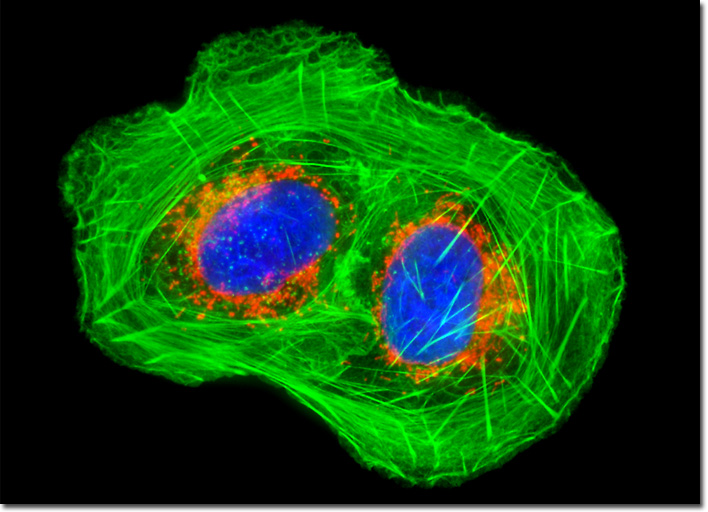
Symptoms of osteosarcoma, which is also known as osteogenic sarcoma, are quite various. However, since the tumor seems to most frequently occur in large bones, such as those of the arms and legs, pain and swelling in these areas may occur. If osteosarcoma develops around the knee, an area that is commonly affected, joint pain and limitation of movement may also accompany the tumor. Unfortunately, osteosarcoma is a very aggressive kind of cancer and the disease may be progressed to an advanced state before symptoms are recognized. Moreover, the tumors metastasize early, regularly resulting in secondary tumors in various areas of the body, including the lungs and the brain, where they can be even more problematic than in bone tissue. In cases where metastases are present, prognosis is significantly worse. A healthy culture of U-2 OS human cancer cells (illustrated above) was stained for mitochondria with MitoTracker CMXRos, a derivative of X-rosamine. After fixation and permeabilization, the cells were labeled with Alexa Fluor 488 conjugated to phalloidin and DAPI, targeting filamentous actin and nuclear DNA, respectively. Images were recorded in grayscale with a QImaging Retiga Fast-EXi camera system coupled to an Olympus BX-51 microscope equipped with bandpass emission fluorescence filter optical blocks provided by Omega Optical. During the processing stage, individual image channels were pseudocolored with RGB values corresponding to each of the fluorophore emission spectral profiles. |
© 1995-2022 by Michael W. Davidson and The Florida State University. All Rights Reserved. No images, graphics, software, scripts, or applets may be reproduced or used in any manner without permission from the copyright holders. Use of this website means you agree to all of the Legal Terms and Conditions set forth by the owners.
This website is maintained by our
|