Fluorescence Digital Image Gallery
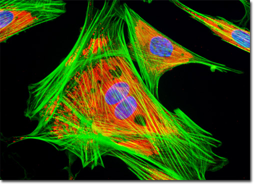
Indian Muntjac Deer Skin Fibroblast Cells
|
Lectins are a specialized class of plant proteins that bind to specific carbohydrate groups attached to proteins or residing in cell membranes. They are commonly utilized in the laboratory to localize glycoproteins and to detect glycol-conjugates in various applications. The lectin known as concanavalin A selectively binds to alpha-mannopyranosyl and alpha-glucopyranosyl residues, which are abundant in the endoplasmic reticulum. When present in acidic solutions, concanavalin A exists as a dimer, although in neutral and alkaline environments the lectin condenses into a tetramer. Concanavalin A has been utilized for a wide range of studies, including investigations of receptor capping in white blood cells, analyses of acrosome reaction incidence in human sperm cells, and examinations focusing upon the lateral diffusion of glycoproteins and viruses in cell membranes. The adherent culture of Indian Muntjac deer skin fibroblasts presented in the digital image above was stained with Texas Red conjugated to the lectin concanavalin A. Alexa Fluor 488 conjugated to phalloidin and DAPI were also used to counterstain the culture, targeting filamentous actin and nuclear DNA, respectively. Images were recorded in grayscale with a QImaging Retiga Fast-EXi camera system coupled to an Olympus BX-51 microscope equipped with bandpass emission fluorescence filter optical blocks provided by Omega Optical. During the processing stage, individual image channels were pseudocolored with RGB values corresponding to each of the fluorophore emission spectral profiles. |
© 1995-2022 by Michael W. Davidson and The Florida State University. All Rights Reserved. No images, graphics, software, scripts, or applets may be reproduced or used in any manner without permission from the copyright holders. Use of this website means you agree to all of the Legal Terms and Conditions set forth by the owners.
This website is maintained by our
|