Fluorescence Digital Image Gallery
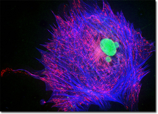
Indian Muntjac Deer Skin Fibroblast Cells
|
MitoTracker probes bind selectively to mitochondria and many are well retained during the fixation of cells with common agents, including methanol, acetone, glutaraldehyde, and paraformaldehyde, enabling their simultaneous use with immunofluorescence applications and counterstains. A mildly thiol-reactive chloromethyl moiety, which is characteristic of these fluorophores, is believed to be responsible for maintaining the association of the dyes with the intracellular mitochondrial network subsequent to fixation. The labeling of mitochondria with MitoTracker stains is relatively simple. Cell cultures are incubated with media in the presence of submicromolar amounts of a selected probe, and the MitoTracker dye passively diffuses across the cellular plasma membrane, accumulating in active mitochondria. Green, orange, and red MitoTracker probes, which vary in their spectral characteristics and fixability, are commercially available. The culture of Indian Muntjac deer skin fibroblast cells presented in the digital image above was labeled for the cytoskeletal filamentous actin network with Alexa Fluor 350 conjugated to phalloidin, and for the cell nucleus with SYTOX Green. Additionally, cellular mitochondria were stained with MitoTracker Red CMXRos, a complex aminated xanthene derivative. Images were recorded in grayscale with a QImaging Retiga Fast-EXi camera system coupled to an Olympus BX-51 microscope equipped with bandpass emission fluorescence filter optical blocks provided by Omega Optical. During the processing stage, individual image channels were pseudocolored with RGB values corresponding to each of the fluorophore emission spectral profiles. |
© 1995-2025 by Michael W. Davidson and The Florida State University. All Rights Reserved. No images, graphics, software, scripts, or applets may be reproduced or used in any manner without permission from the copyright holders. Use of this website means you agree to all of the Legal Terms and Conditions set forth by the owners.
This website is maintained by our
|