Fluorescence Digital Image Gallery
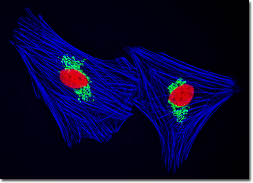
Indian Muntjac Deer Skin Fibroblast Cells
|
The stacks of cisternae that comprise the Golgi apparatus exhibit a distinct polarity. The two opposing faces of a stack are known as the cis face and the trans face. The cis face is generally positioned in the cell so that it is near the endoplasmic reticulum, an arrangement that readily enables this entry face to accept transport vesicles from the organelle. Thus, the cis face is often considered to be the receiving department of the Golgi complex. Correspondingly, the trans or exit face is commonly deemed the shipping department of the organelle since new vesicles bud from it and are dispatched to other locations in the cell or to the cell's surface. Due to this function of the Golgi stack, it is not surprising that the Golgi apparatus is most extensive in secretory cells, such as goblet cells of the small intestine. The culture of Muntjac cells illustrated above was triple-labeled using double immunofluorescence and a phallotoxin. Nuclei were visualized with mouse anti-histones (core) primary antibodies, while the Golgi complex was stained with rabbit anti-giantin antibodies. Secondary antibodies were goat anti-mouse and anti-rabbit conjugated to Texas Red and Oregon Green 488, respectively to produce red nuclei and green Golgi cisternae. The filamentous actin network was counterstained with Alexa Fluor 350 conjugated to phalloidin. Images were recorded in grayscale with a QImaging Retiga Fast-EXi camera system coupled to an Olympus BX-51 microscope equipped with bandpass emission fluorescence filter optical blocks provided by Omega Optical. During the processing stage, individual image channels were pseudocolored with RGB values corresponding to each of the fluorophore emission spectral profiles. |
© 1995-2022 by Michael W. Davidson and The Florida State University. All Rights Reserved. No images, graphics, software, scripts, or applets may be reproduced or used in any manner without permission from the copyright holders. Use of this website means you agree to all of the Legal Terms and Conditions set forth by the owners.
This website is maintained by our
|