Fluorescence Digital Image Gallery
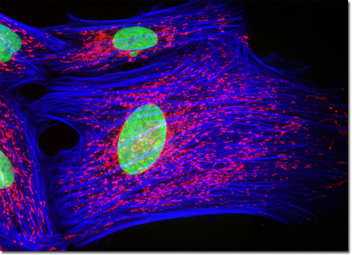
Indian Muntjac Deer Skin Fibroblast Cells
|
Phallotoxins are a group of potent hepatotoxins found in many mushroom species from the genus Amanita. The phallotoxins known as phalloidin and phallacidin are both isolated specifically from Amanita phalloides, a member that is more commonly known as the death cap. Often used in cell labeling applications, the bicyclic peptides differ by only two amino acid residues and can, therefore, frequently be utilized interchangeably. Phalloidin and phallacidin specifically target filamentous actin, binding competitively to the same sites on the macromolecule. It is also important to note that the two phallotoxins contain an atypical thioether connection between a cysteine and tryptophan residue, which structurally creates an inner ring. At unusually high pH levels, this thioether linkage is broken and, consequently, the high affinity of phalloidin and phallacidin for F-actin is lost. The Indian Muntjac fibroblast cell culture appearing in the digital image above was labeled with Alexa Fluor 350 conjugated to phalloidin, targeting the F-actin cytoskeletal network. The cells were also stained with MitoTracker Red CMXRos and SYTOX Green, which preferentially bind with intracellular mitochondria and DNA in the cell nucleus, respectively. Images were recorded in grayscale with a QImaging Retiga Fast-EXi camera system coupled to an Olympus BX-51 microscope equipped with bandpass emission fluorescence filter optical blocks provided by Omega Optical. During the processing stage, individual image channels were pseudocolored with RGB values corresponding to each of the fluorophore emission spectral profiles. |
© 1995-2022 by Michael W. Davidson and The Florida State University. All Rights Reserved. No images, graphics, software, scripts, or applets may be reproduced or used in any manner without permission from the copyright holders. Use of this website means you agree to all of the Legal Terms and Conditions set forth by the owners.
This website is maintained by our
|