Fluorescence Digital Image Gallery
Indian Muntjac Deer Skin Fibroblast Cells
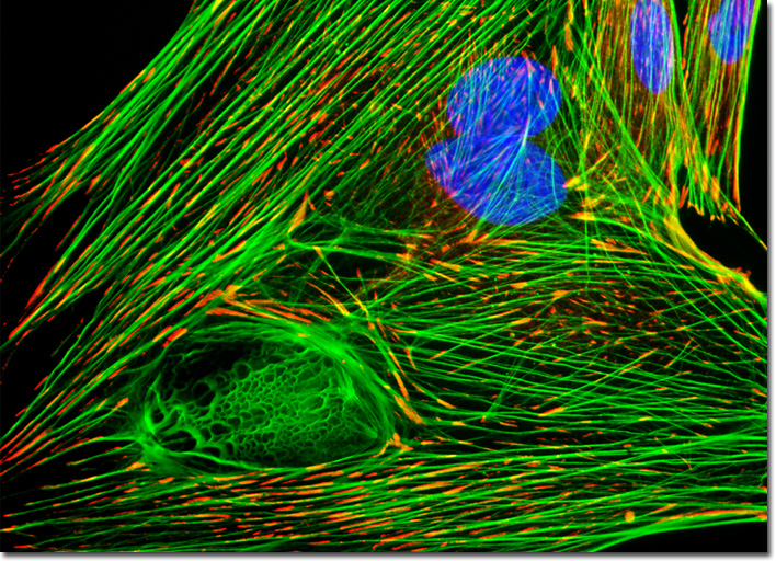
|
The popular Hoechst dyes, which are a class of commonly used fluorescent nucleic acid probes (particularly Hoechst 33258, Hoechst 33342, and Hoechst 34580), are produced by a global corporate group headquartered in Germany. These dyes can be excited with the ultraviolet and violet spectral lines of the helium-cadmium, argon-ion, and blue diode lasers, as well as by most standard fluorescence arc-discharge excitation sources. They exhibit relatively large Stokes shifts, which render the probes appropriate for multicolor labeling experiments and a number of other applications, including cell cycle and apoptosis investigations. Moreover, Hoechst 33258 is selectively toxic to malaria parasites and has, therefore, been used in the flow-cytometric screening of blood samples for the organisms. In general, Hoechst dyes demonstrate a wide range of sequence-dependent DNA affinities and reportedly bind with sufficient strength to certain nucleotide sequences that they are often capable of displacing tightly bound DNA intercalators when applied in high concentrations. The culture of Indian Muntjac deer skin fibroblast cells featured in the digital image above was immunofluorescently labeled with primary anti-vinculin mouse monoclonal antibodies followed by goat anti-mouse Fab fragments conjugated to the cyanine dye, Cy3. Vinculin is a protein associated with the cytoplasmic face of focal adhesions. In addition, the fixed and permeabilized culture was counterstained for DNA with the probe Hoechst 33258, and for the cytoskeletal filamentous actin network with Alexa Fluor 488 conjugated to phalloidin. Images were recorded in grayscale with a QImaging Retiga Fast-EXi camera system coupled to an Olympus BX-51 microscope equipped with bandpass emission fluorescence filter optical blocks provided by Omega Optical. During the processing stage, individual image channels were pseudocolored with RGB values corresponding to each of the fluorophore emission spectral profiles. |
© 1995-2025 by Michael W. Davidson and The Florida State University. All Rights Reserved. No images, graphics, software, scripts, or applets may be reproduced or used in any manner without permission from the copyright holders. Use of this website means you agree to all of the Legal Terms and Conditions set forth by the owners.
This website is maintained by our
|