Fluorescence Digital Image Gallery
Indian Muntjac Deer Skin Fibroblast Cells
|
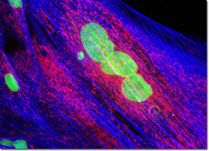
Several different classes of proprietary cyanine dyes are commercially available, including several that are ultra-sensitive, chemically reactive, conjugated, cell-permeant, and cell-impermeant. This latter class of dyes, which only penetrate the compromised membranes characteristic of dead cells, includes the popular TOTO, TO-PRO, and SYTOX families of fluorophores. The specific spectral profiles and binding affinities of these dyes vary across a wide range and are, therefore, utilized for an array of applications. The SYTOX family, which includes dyes that fluoresce green, blue, and orange, are considered to be particularly useful for dead cell assays and as DNA counterstains for nuclear and chromosome labeling in fixed and permeabilized preparations. Several of the SYTOX dyes can be employed in combination with traditional cell-permeant probes, such as DAPI and Hoechst 33342, for two-color visualization of live and dead cells. In addition, SYTOX nucleic acid stains are an excellent choice in multi-labeling experiments with other fluorophores that target cytoplasmic organelles, the cytoskeleton, microfilaments, and specific enzymes and proteins. The culture of Indian Muntjac deer skin fibroblast cells presented in the digital image above was labeled for filamentous actin with Alexa Fluor 350 conjugated to phalloidin, and for the cellular mitochondrial network with MitoTracker Red CMXRos. SYTOX Green was also used to counterstain the cells, targeting nuclear DNA. Images were recorded in grayscale with a QImaging Retiga Fast-EXi camera system coupled to an Olympus BX-51 microscope equipped with bandpass emission fluorescence filter optical blocks provided by Omega Optical. During the processing stage, individual image channels were pseudocolored with RGB values corresponding to each of the fluorophore emission spectral profiles. |
© 1995-2025 by Michael W. Davidson and The Florida State University. All Rights Reserved. No images, graphics, software, scripts, or applets may be reproduced or used in any manner without permission from the copyright holders. Use of this website means you agree to all of the Legal Terms and Conditions set forth by the owners.
This website is maintained by our
|