Fluorescence Digital Image Gallery
Human Fetal Lung Fibroblast Cells (MRC-5)
|
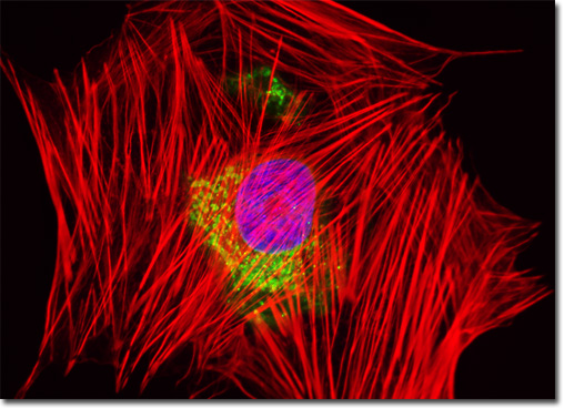
The cytoskeleton of a cell is a lattice located in the cytoplasmic matrix. The structural scaffolding, which guides and moves intracellular material in addition to providing support, is comprised of both tubular and fibrillar elements. The fibrillar components are largely comprised of actin, a protein that exists in both globular (G-actin) and filamentous (F-actin) forms. In nonmuscle cells, actin comprises approximately 15 percent of the total protein content. Of this amount, about half exists in the polymeric filamentous form. Actin molecules present in a wide variety of species differ very little in their amino acid sequences, which is suggestive of the highly conserved nature of the protein. The single human fetal lung fibroblast (MRC-5) cell that is featured in the digital image above was resident in a culture that was stained with Alexa Fluor 568 conjugated to phalloidin, which binds to the cytoskeletal filamentous actin network. The cells were subsequently stained with Hoechst 33258 (targeting DNA in the cell nucleus) and the lectin wheat germ agglutinin conjugated to Oregon Green 488 (targeting glycoproteins in the Golgi complex). Images were recorded in grayscale with a QImaging Retiga Fast-EXi camera system coupled to an Olympus BX-51 microscope equipped with bandpass emission fluorescence filter optical blocks provided by Omega Optical. During the processing stage, individual image channels were pseudocolored with RGB values corresponding to each of the fluorophore emission spectral profiles. |
© 1995-2025 by Michael W. Davidson and The Florida State University. All Rights Reserved. No images, graphics, software, scripts, or applets may be reproduced or used in any manner without permission from the copyright holders. Use of this website means you agree to all of the Legal Terms and Conditions set forth by the owners.
This website is maintained by our
|