Fluorescence Digital Image Gallery
Human Fetal Lung Fibroblast Cells (MRC-5)
|
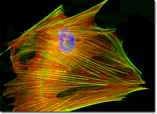
Human lung development is not completed until after birth. The lung of a newborn features only a small fraction of the number of alveoli found in a mature adult lung. Thus, alveolar formation continues for some time postnatally, most experts believing that the process completes at around two years of age. Yet, even when formation of alveoli is concluded, the lung is still not fully mature. At this stage, a double capillary network remains in the interalveolar septa, although adult lungs feature a single capillary network. Therefore, an intensive reorganization must still take place for development of the lung to be considered complete. Generally, this process is finished by the time a child is five years old. Following this final step of development, the lung can begin the normal course of growth. A culture of human lung fetal fibroblast cells (illustrated above) was stained with Alexa Fluor 488 conjugated to phalloidin, which targets cytoskeletal filamentous actin, and Hoechst 33258, which binds to DNA in cell nuclei. The MRC-5 cells were also immunofluorescently labeled with anti-tubulin mouse monoclonal primary antibodies followed by goat anti-mouse Fab fragments conjugated to Cy3, targeting the intracellular microtubular network. Images were recorded in grayscale with a QImaging Retiga Fast-EXi camera system coupled to an Olympus BX-51 microscope equipped with bandpass emission fluorescence filter optical blocks provided by Omega Optical. During the processing stage, individual image channels were pseudocolored with RGB values corresponding to each of the fluorophore emission spectral profiles. |
© 1995-2022 by Michael W. Davidson and The Florida State University. All Rights Reserved. No images, graphics, software, scripts, or applets may be reproduced or used in any manner without permission from the copyright holders. Use of this website means you agree to all of the Legal Terms and Conditions set forth by the owners.
This website is maintained by our
|