Fluorescence Digital Image Gallery
Human Fetal Lung Fibroblast Cells (MRC-5)
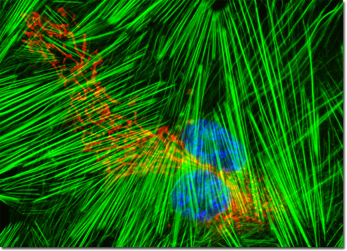
|
The development of the human lung takes place in several stages. In a fetus, such as the one that served as the source for the MRC-5 cell line, the lung primordium is established after early embryogenesis. The epithelial sections of the organ are derivatives of the gut, whereas the tissues and blood vessels that surround these areas originate from the mesoderm. When the lung begins to appear like a gland subsequent to a series of dichotomous divisions, it is said to be in the pseudoglandular stage, a phase that is also characterized by the development of vascular connections and the area of tubules that mark the future region of gas-exchange activity. The next step in fetal lung development is the canalicular stage, in which potential airspaces expand, the cuboidal epithelium differentiates into different types of cells, and a thin latent air-blood barrier is formed. The final stage of lung development in the fetus is called the saccular stage. At this point, additional airways and saccules are formed. Near the end of the saccular stage, alveoli begin to develop through septation of the saccules. The culture of human fetal lung fibroblast (MRC-5) cells presented in the digital image above was labeled with wheat germ agglutinin, a lectin that targets glycoproteins in the Golgi network, conjugated to Texas Red-X. In addition, the cells were labeled for the cytoskeletal F-actin network and DNA with Alexa Fluor 488 conjugated to phalloidin and DAPI, respectively. Images were recorded in grayscale with a QImaging Retiga Fast-EXi camera system coupled to an Olympus BX-51 microscope equipped with bandpass emission fluorescence filter optical blocks provided by Omega Optical. During the processing stage, individual image channels were pseudocolored with RGB values corresponding to each of the fluorophore emission spectral profiles. |
© 1995-2025 by Michael W. Davidson and The Florida State University. All Rights Reserved. No images, graphics, software, scripts, or applets may be reproduced or used in any manner without permission from the copyright holders. Use of this website means you agree to all of the Legal Terms and Conditions set forth by the owners.
This website is maintained by our
|