Fluorescence Digital Image Gallery
Madin-Darby Ovine Kidney Epithelial Cells (MDOK)
|
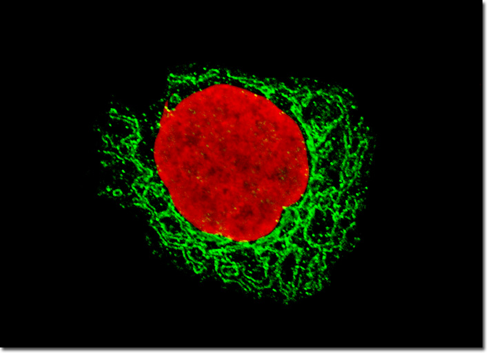
Histones are proteins that occur in the nuclei of most cells in ionic combination with DNA. Nucleosome is the term for a histone molecule bound to a segment of DNA, and nucleosomes are subunits of chromatin, the readily stainable substance that condenses to form chromosomes during cell division. Histones were discovered in the late 1800s by Albrecht Kossel, a German doctor who received the Nobel Prize in Physiology or Medicine in 1910 for his research in the field of cell biology. However, much of the modern understanding of the water-soluble, alkaline proteins was not developed until the latter half of the twentieth century. Indeed, histones were generally assumed to serve primarily as a type of packing for DNA to coil around for many years, but today the proteins are widely considered to be involved in gene regulation, the physical and functional alterations that occur in chromatin during cell division, and the transcription of DNA. The Madin-Darby ovine kidney cell culture featured in the digital image above was fixed, permeabilized, washed, and blocked with 10-percent normal goat serum in phosphate-buffered saline prior to immunofluorescent labeling with rabbit primary antibodies to giantin, a protein resident in the Golgi complex of mammalian cells. The culture was subsequently stained with a mixture of secondary antibodies conjugated to Alexa Fluor 488. In addition, histones were immunofluorescently labeled with primary anti-histone mouse monoclonal antibodies followed by goat anti-mouse Fab heavy and light chain fragments conjugated to Texas Red. Images were recorded with a FluoView FV300 laser scanning confocal microscope using argon-ion and helium-neon laser systems. |
© 1995-2022 by Michael W. Davidson and The Florida State University. All Rights Reserved. No images, graphics, software, scripts, or applets may be reproduced or used in any manner without permission from the copyright holders. Use of this website means you agree to all of the Legal Terms and Conditions set forth by the owners.
This website is maintained by our
|