Fluorescence Digital Image Gallery
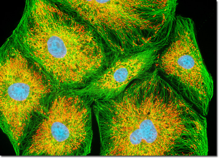
Madin-Darby Ovine Kidney Epithelial Cells (MDOK)
|
Most eukaryotic cells exhibit three different varieties of cytoskeletal filaments, each of which functions in a characteristic manner. The long, hollow filaments known as microtubules are primarily responsible for regulating the transport of molecules between intracellular compartments and governing the positioning of organelles bound by membranes, such as the mitochondria illustrated in the digital image above. A microtubule exhibits an outer diameter of approximately 25 nanometers and is significantly more rigid than an actin filament. In a typical animal cell, microtubules are nucleated around a small region of cytoplasm bordering the nucleus of the cell called a microtubule-organizing center (MTOC), or centrosome. From the MTOC, the filaments extend out in an astral-like configuration, with their minus ends embedded in the centrosome and their plus ends situated towards the periphery of the cell. A specialized variety of tubulin (gamma-tubulin) apparently plays a key role in the organization of microtubules, forming ring-shaped complexes with proteins that are widely supposed to act as templates for the nucleation of microtubules that have grown to contain the 13 protafilaments characteristic of the structures. In order to visualize the interrelationship between the microtubule and mitochondrial networks in the MDOK culture illustrated above, a rapidly growing adherent culture was stained with MitoTracker Red CMXRos, fixed, permeabilized, and then treated with mouse primary antibodies to tubulin. The culture was subsequently labeled with goat anti-mouse Fab antibody fragments conjugated to fluorescein and counterstained with Hoechst 33258 (targeting DNA in the nucleus). Images were recorded in grayscale with a QImaging Retiga Fast-EXi camera system coupled to an Olympus BX-51 microscope equipped with bandpass emission fluorescence filter optical blocks provided by Omega Optical. During the processing stage, individual image channels were pseudocolored with RGB values corresponding to each of the fluorophore emission spectral profiles. |
© 1995-2022 by Michael W. Davidson and The Florida State University. All Rights Reserved. No images, graphics, software, scripts, or applets may be reproduced or used in any manner without permission from the copyright holders. Use of this website means you agree to all of the Legal Terms and Conditions set forth by the owners.
This website is maintained by our
|