Fluorescence Digital Image Gallery
Madin-Darby Canine Kidney Epithelial Cells (MDCK)
|
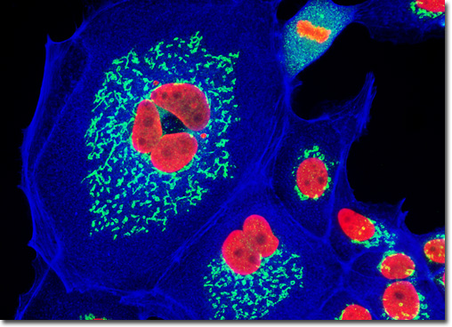
In order to label the Golgi domain in mammalian cells with immunofluorescent probes, a candidate protein residing somewhere in the complex must be identified and targeted with a primary mono or polyclonal antibody. Giantin is a membrane-bound glycoprotein, which is resident in the cis and medial Golgi regions, and contains an elongated rod-like cytoplasmic domain that is at least 350 kiloDaltons in size. This highly conserved protein may be involved in intercisternal cross-bridge structures that are suspected to play a role in Golgi cisternae stacking. Polyclonal antibodies raised against the N-terminal 469 peptide residues of giantin react with a number of mammalian species, and monoclonal antibodies have been produced that detect the protein in avians, as well. The culture of Madin-Darby canine kidney epithelial cells appearing in the digital image above was fixed with paraformaldehyde, permeabilized, and treated with a mixture of rabbit (anti-giantin) and mouse (anti-histones; pan) primary antibodies, followed by secondary antibodies conjugated to Alexa Fluor 488 and Texas Red, respectively. The secondary antibody cocktail also contained Alexa Fluor 350 conjugated to phalloidin, designed to simultaneously target the filamentous actin network. Images were recorded in grayscale with a QImaging Retiga Fast-EXi camera system coupled to an Olympus BX-51 microscope equipped with bandpass emission fluorescence filter optical blocks provided by Omega Optical. During the processing stage, individual image channels were pseudocolored with RGB values corresponding to each of the fluorophore emission spectral profiles. |
© 1995-2022 by Michael W. Davidson and The Florida State University. All Rights Reserved. No images, graphics, software, scripts, or applets may be reproduced or used in any manner without permission from the copyright holders. Use of this website means you agree to all of the Legal Terms and Conditions set forth by the owners.
This website is maintained by our
|