Fluorescence Digital Image Gallery
Madin-Darby Canine Kidney Epithelial Cells (MDCK Line)
Since most epithelial tissues cover or line surfaces, they are often exposed to mechanical abrasion. Thus, epithelial cells must be able to readily replace themselves if necessary. Indeed, the cells are fully capable of cellular division, a process that occurs in epithelia rather rapidly. For instance, the epithelium that lines the small intestine is believed to be fully replaced with newly divided cells at least every six days. As new cells are created, old cells are sloughed. The outermost epithelium of the body, the skin, is another good example of the proliferation capabilities of epithelial cells. New skin cells are produced on an almost continuous basis in the innermost layers of the dermis in order to maintain the integrity of the outer layers, the cells of which persistently flake away. The adherent culture of Madin-Darby canine kidney cells presented in the digital image above was labeled for the intracellular mitochondrial network and for filamentous actin with MitoTracker Red CMXRos and Alexa Fluor 488 conjugated to phalloidin, respectively. The ultraviolet-absorbing probe DAPI was utilized to counterstain DNA. Images were recorded in grayscale with a QImaging Retiga Fast-EXi camera system coupled to an Olympus BX-51 microscope equipped with bandpass emission fluorescence filter optical blocks provided by Omega Optical. During the processing stage, individual image channels were pseudocolored with RGB values corresponding to each of the fluorophore emission spectral profiles.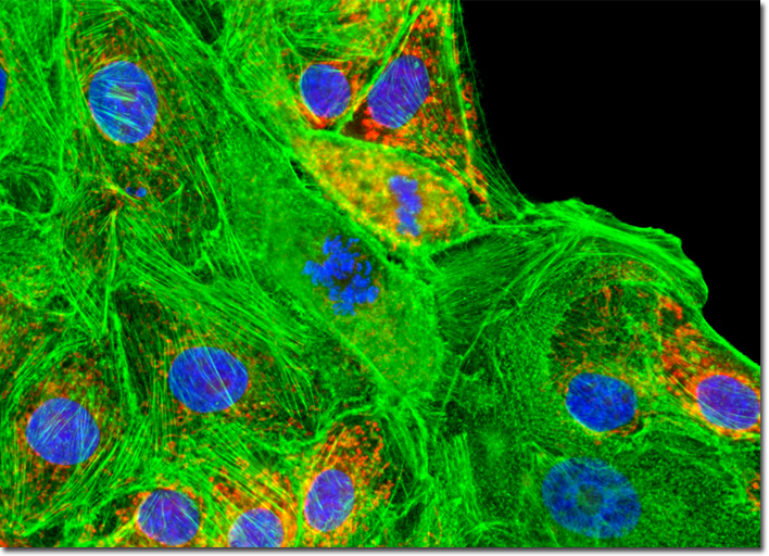
© 1995-2022 by Michael W. Davidson and The Florida State University. All Rights Reserved. No images, graphics, software, scripts, or applets may be reproduced or used in any manner without permission from the copyright holders. Use of this website means you agree to all of the Legal Terms and Conditions set forth by the owners.
This website is maintained by our
|