Fluorescence Digital Image Gallery
Human Cervical Adenocarcinoma Cells (HeLa)
|
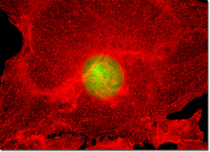
According to some reports, incidence of adenocarcinoma of the cervix is rising, although the incidence of other cervical cancers, such as those of the squamous type, is decreasing. Moreover, this increase in incidence seems to be most pronounced in young women. In fact, findings indicate that the incidence of adenocarcinoma of the cervix in the United States more than doubled between the early 1970s and the mid 1980s among women less than 35 years old. Some scientists have suggested that this significant change may be a result of the widespread use of oral contraceptives that began soon after they were introduced in the early 1960s. In the few investigative studies that have been carried out in this area, the data obtained seems to support this hypothesis, women who have taken oral contraceptives for extensive periods of time exhibiting a significantly higher risk of adenocarcinoma of the cervix than those who have never taken them. The culture of HeLa carcinoma cells featured in the digital image above was transfected with a pEGFP-Nucleus plasmid subcellular localization vector. Plasmid pEYFP-Nucleus vector gene product expression in various cell types occurs due to the efficient intracellular translation of a fusion nucleotide sequence combining the enhanced green fluorescent protein domain with three tandem repeats of the nuclear localization signal (NLS) from simian virus 40 (SV40) large T-antigen. In addition, the cells were labeled with Alexa Fluor 568 conjugated to phalloidin in order to target the cytoskeletal filamentous actin network. Images were recorded in grayscale with a QImaging Retiga Fast-EXi camera system coupled to an Olympus BX-51 microscope equipped with bandpass emission fluorescence filter optical blocks provided by Omega Optical. During the processing stage, individual image channels were pseudocolored with RGB values corresponding to each of the fluorophore emission spectral profiles. |
© 1995-2025 by Michael W. Davidson and The Florida State University. All Rights Reserved. No images, graphics, software, scripts, or applets may be reproduced or used in any manner without permission from the copyright holders. Use of this website means you agree to all of the Legal Terms and Conditions set forth by the owners.
This website is maintained by our
|