
Fluorescence Digital Image Gallery
Mouse Hemangioendothelioma Endothelial Cells (EOMA Line)
The EOMA cell line was derived in 1980 from a mixed hemangioendothelioma present in an adult mouse (Mus musculus). Hemangioendothelioma is the term utilized to describe a varied group of vascular tumors that usually appear as red or blue nodules and tend to behave biologically in a manner that can be classified as falling between a benign hemangioma and malignant angiosarcoma.
EOMA cells are tumorigenic in syngeneic mice and exhibit characteristic endothelial cell properties. The cells synthesize a number of cellular products including angiotensin-converting enzyme (ACE), thrombospondin, Cathepsin L, endostatin, and interleukin-6. Surface receptors for acetylated low-density lipoprotein are expressed by EOMA cells, as is the vascular addressin, a tissue-specific endothelial cell adhesion molecule identified by antibody MECA-99.
Dilated and congested veins with inactive endothelial cell nuclei and occasional thrombi or phleboliths are typical of hemangioendothelioma. Solid sheets of epitheloid or spindle-shaped mesenchymal cells are also often present in the affected region, where they intermix with the problematic blood vessels. Approximately one-tenth of the cases of the condition are associated with other medical problems, including Maffucci's syndrome, lymphedema, and premature onset varicose veins. The prognosis for the condition varies, some of the tumors being much more destructive and likely to recur or metastasize than others.
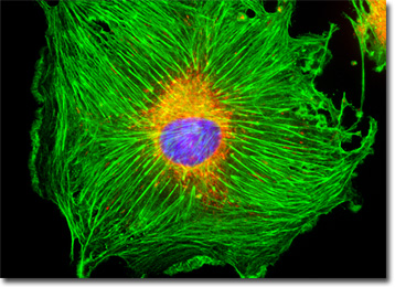
The EOMA cell culture featured in the digital image above was labeled with MitoTracker Red CMXRos, Alexa Fluor 488 conjugated to phalloidin, and DAPI, targeting the intracellular mitochondrial network, cytoskeletal F-actin, and nuclear DNA, respectively. Images were recorded in grayscale with a QImaging Retiga Fast-EXi camera system coupled to an Olympus BX-51 microscope equipped with bandpass emission fluorescence filter optical blocks provided by Omega Optical. During the processing stage, individual image channels were pseudocolored with RGB values corresponding to each of the fluorophore emission spectral profiles.
BACK TO THE CULTURED CELLS FLUORESCENCE GALLERY
BACK TO THE FLUORESCENCE GALLERY