Fluorescence Digital Image Gallery
Rat Jejunum Myenteric Plexus Enteroglial Cells (EGC/PK060399egfr)
|
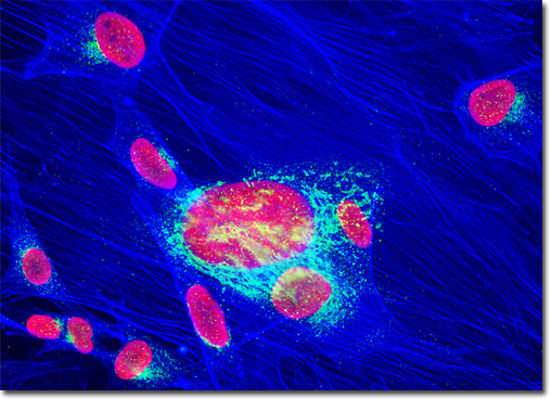
DNA packaging in eukaryotic cells is a multifarious task due to the fact that the length of the molecule greatly exceeds the diameter of the nucleus in which it is housed (when lined up end-to end, a human cell contains approximately 2 meters of DNA). Moreover, chromosomes are dynamic structures and cells must be able to readily access specific DNA sequences when needed in order to accomplish activities such as replication and repair. Albrecht Kossel, a German scientist who was awarded the Nobel Prize in Medicine or Physiology for his work in cellular biology in 1910, discovered a group of specialized proteins that are intricately involved in the achievement of this monumental task in the 1880s. Dubbed histones, these proteins occur in massive quantities in eukaryotic cells, where they account for the formation of nucleosomes, the most rudimentary unit of chromosome organization. When subjected to treatments that incite its partial unpacking and then examined under an electron microscope, chromatin exhibits the appearance of beads on a string. The string-like areas of the complex are sequences of linker DNA and the beads are nucleosome core particles, each comprised of DNA coiled around a histone-based protein core. The proximity of the Golgi complex and nuclei in the rat enteroglial cell culture illustrated above was probed in a double immunofluorescence experiment with mouse anti-histones (pan) and rabbit anti-giantin primary antibodies. The antibody targets were visualized with goat secondary antibodies conjugated to Texas Red and Alexa Fluor 488, respectively, while the actin cytoskeletal framework was labeled with Alexa Fluor 350 conjugated to phalloidin. Images were recorded in grayscale with a QImaging Retiga Fast-EXi camera system coupled to an Olympus BX-51 microscope equipped with bandpass emission fluorescence filter optical blocks provided by Omega Optical. During the processing stage, individual image channels were pseudocolored with RGB values corresponding to each of the fluorophore emission spectral profiles. |
© 1995-2022 by Michael W. Davidson and The Florida State University. All Rights Reserved. No images, graphics, software, scripts, or applets may be reproduced or used in any manner without permission from the copyright holders. Use of this website means you agree to all of the Legal Terms and Conditions set forth by the owners.
This website is maintained by our
|