Fluorescence Digital Image Gallery
Rat Jejunum Myenteric Plexus Enteroglial Cells (EGC/PK060399egfr)
|
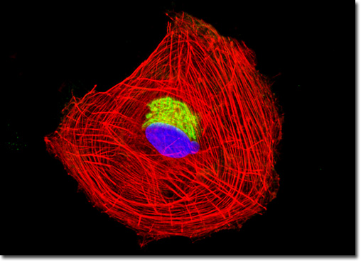
A number of different substances can be found in the enteroglial cells of the myenteric plexus that appear to play important roles in the activity of the nerve center. For instance, the heavily studied neuropeptide known as substance P is present in the myenteric plexus in significant levels and may help facilitate the production of saliva, smooth muscle contractions, and other tissue responses. Both endorphins and enkephalins, which are peptide hormones with opiate qualities, are also found in the myenteric plexus. There is some uncertainty regarding the exact roles of these substances, but it is often suggested that the enkephalins are involved in the regulation of smooth muscle activity, while the endorphins may help regulate the secretion of other peptides by the digestive system’s endocrine cells. Other substances have also been identified in this part of the enteric nervous system, but further studies are necessary to more clearly delineate their respective functions. The culture of rat enteroglial cells presented in the digital image above was labeled with Oregon Green 488 conjugated to the lectin wheat germ agglutinin. Fluorescent wheat germ agglutinin conjugates are often used as probes for the Golgi network in mammalian cultures. The cells were also stained with Alexa Fluor 568 conjugated to phalloidin and DAPI, which target F-actin and DNA, respectively. Images were recorded in grayscale with a QImaging Retiga Fast-EXi camera system coupled to an Olympus BX-51 microscope equipped with bandpass emission fluorescence filter optical blocks provided by Omega Optical. During the processing stage, individual image channels were pseudocolored with RGB values corresponding to each of the fluorophore emission spectral profiles. |
© 1995-2022 by Michael W. Davidson and The Florida State University. All Rights Reserved. No images, graphics, software, scripts, or applets may be reproduced or used in any manner without permission from the copyright holders. Use of this website means you agree to all of the Legal Terms and Conditions set forth by the owners.
This website is maintained by our
|