Fluorescence Digital Image Gallery
Rat Jejunum Myenteric Plexus Enteroglial Cells (EGC/PK060399egfr)
|
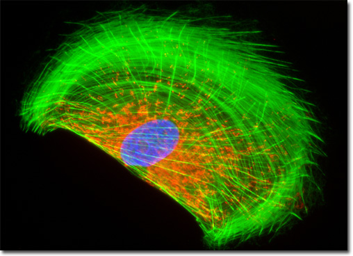
The enteric nervous system is composed of networked nerve fibers that cooperatively control the movement of the gastrointestinal tract. Though many details about the functioning of this system have not yet been fully realized, it is considered to be chiefly comprised of two different nerve centers: the myenteric plexus and the submucosal plexus. The myenteric plexus, which is the collection of nerves that served as the source for the EGC/PK060399egfr cell line, can be found in the lower esophagus, stomach, and intestines lying between the inner circular muscle layer and the outer longitudinal muscle layer. Scientists generally agree that it is this plexus, which receives input from the vagus nerve, that regulates the peristaltic waves that move food through the digestive system as well as the muscular contractions that result in fixed churning. The submucosal plexus, however, is buried in the submucosal tissue of the gastrointestinal tract and is apparently involved in the production of glandular secretions and the regulation of local blood flow and electrolyte and water transport. The mitochondria present in the culture of rat enteroglial cells featured in the digital image above were labeled with MitoTracker Red CMXRos, a derivative of X-rosamine. In addition, the culture was labeled for the cytoskeletal F-actin network and DNA in the cell nucleus with Alexa Fluor 488 conjugated to phalloidin and DAPI, respectively. Images were recorded in grayscale with a QImaging Retiga Fast-EXi camera system coupled to an Olympus BX-51 microscope equipped with bandpass emission fluorescence filter optical blocks provided by Omega Optical. During the processing stage, individual image channels were pseudocolored with RGB values corresponding to each of the fluorophore emission spectral profiles. |
© 1995-2022 by Michael W. Davidson and The Florida State University. All Rights Reserved. No images, graphics, software, scripts, or applets may be reproduced or used in any manner without permission from the copyright holders. Use of this website means you agree to all of the Legal Terms and Conditions set forth by the owners.
This website is maintained by our
|