Fluorescence Digital Image Gallery
Transformed African Green Monkey Kidney Fibroblast Cells (COS-7)
|
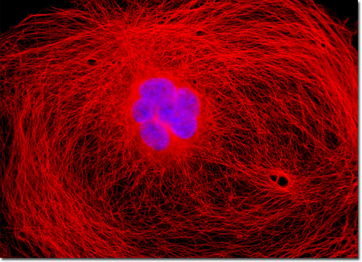
The choice of fluorescent probes for confocal and fluorescence microscopy must address the specific capabilities of the instrument to excite and detect fluorescence emission in the wavelength regions made available by the laser systems and detectors. Although the current lasers used in confocal and fluorescence microscopy produce discrete lines in the ultraviolet, visible, and near-infrared portions of the spectrum, the location of these spectral lines does not always coincide with absorption maxima of popular fluorophores. In fact, it is not necessary for the laser spectral line to correspond exactly with the fluorophore wavelength of maximum absorption, but the intensity of fluorescence emission is regulated by the fluorophore extinction coefficient at the excitation wavelength. The most popular lasers for confocal and fluorescence microscopy are air-cooled argon and krypton-argon ion lasers, the new blue diode lasers, and a variety of helium-neon systems. Collectively, these lasers are capable of providing excitation at ten to twelve specific wavelengths between 400 and 650 nanometers. The African green monkey kidney (COS-7) fibroblast cell appearing in the digital image above was resident in a culture that was immunofluorescently labeled with primary anti-tubulin mouse monoclonal antibodies followed by goat anti-mouse Fab fragments conjugated to Cy3, targeting the extensive cellular microtubular network. The culture was also stained for DNA with the ultraviolet-absorbing probe DAPI. Images were recorded in grayscale with a QImaging Retiga Fast-EXi camera system coupled to an Olympus BX-51 microscope equipped with bandpass emission fluorescence filter optical blocks provided by Omega Optical. During the processing stage, individual image channels were pseudocolored with RGB values corresponding to each of the fluorophore emission spectral profiles. |
© 1995-2022 by Michael W. Davidson and The Florida State University. All Rights Reserved. No images, graphics, software, scripts, or applets may be reproduced or used in any manner without permission from the copyright holders. Use of this website means you agree to all of the Legal Terms and Conditions set forth by the owners.
This website is maintained by our
|