Fluorescence Digital Image Gallery
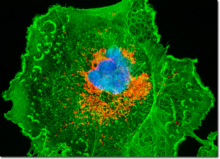
Transformed African Green Monkey Kidney Fibroblast Cells (COS-1)
|
In recent years, a concerted effort has been made by the scientific community to determine whether exposure to SV40 through polio vaccinations is responsible for causing cancer or other health problem in humans. This important endeavor was sparked by Michele Carbone, an Italian pathologist who first detected SV40 DNA in tumors in the early 1990s. Before his work, the research community had widely assumed the vaccine contaminant to be harmless to humans, chiefly based on studies carried out in the 1960s soon after the discovery of the virus. Since that time, SV40 came to be heavily utilized in laboratory studies of cancer because it so easily produces a variety of tumors in laboratory animals, yet little research in the subsequent decades was concerned with what effect the virus might have on people. Due to Carbone’s groundbreaking studies, however, researchers are examining SV40 in a new light. The COS-1 African green monkey kidney fibroblast illustrated in the digital image above was resident in a culture labeled with MitoTracker Red CMXRos (yielding red emission) and Alexa Fluor 488 (green emission) conjugated to phalloidin in order to target mitochondria and the cytoskeletal F-actin network, respectively. In addition, the nucleus was counterstained with the DNA-selective bisbenzimide dye, Hoechst 33258 (blue emission). Images were recorded in grayscale with a QImaging Retiga Fast-EXi camera system coupled to an Olympus BX-51 microscope equipped with bandpass emission fluorescence filter optical blocks provided by Omega Optical. During the processing stage, individual image channels were pseudocolored with RGB values corresponding to each of the fluorophore emission spectral profiles. |
© 1995-2022 by Michael W. Davidson and The Florida State University. All Rights Reserved. No images, graphics, software, scripts, or applets may be reproduced or used in any manner without permission from the copyright holders. Use of this website means you agree to all of the Legal Terms and Conditions set forth by the owners.
This website is maintained by our
|