Fluorescence Digital Image Gallery
Transformed African Green Monkey Kidney Fibroblast Cells (COS-1 Line)
The transformed COS-1 cell line was derived from the CV-1 fibroblast line, which was initiated in March, 1964 by F. C. Jensen and colleagues from the normal kidney of an adult African green monkey. Developed by Yakov Gluzman, the COS-1 cell line differs from CV-1 due to transformation of the earlier line with an origin defective mutant of simian virus 40 (SV40) that codes for wild type T-antigen.

Two other transformed lines, COS-3 and COS-7, were also established by Gluzman, but COS-1 cells are unique in the fact that they contain a single integrated copy of the complete early region of SV40 DNA. All three of the lines, however, fully support the lytic growth of SV40 (tsA209 strain) at 40 degrees Celsius and the replication of SV40 mutants with deletions in the early region.
The kidney cells of African green monkeys, as well as those of rhesus monkeys, have been widely used to produce polio vaccines since the 1950s, and evidence suggests that many of the early vaccines produced were contaminated with various simian viruses. SV40, for instance, had already been introduced to a significant portion of the human population through tainted vaccines by the time of its discovery in 1960. A member of the Papovaviridae family of viruses, SV40 typically invades tissues, multiplies, and then kills cells as the new viruses emerge in certain simian hosts. However, when SV40 infects some mammals, the virus is often unable to reproduce, but may form T-antigen and transform host cells into cancer cells. Indeed, studies have shown that the virus induces tumors in rodents and mounting evidence suggests that SV40 may be associated with some types of human cancer, such as mesothelioma.
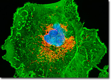
The COS-1 African green monkey kidney fibroblast illustrated in the digital image above was resident in a culture labeled with MitoTracker Red CMXRos (yielding red emission) and Alexa Fluor 488 (green emission) conjugated to phalloidin in order to target mitochondria and the cytoskeletal F-actin network, respectively. In addition, the nucleus was counterstained with the DNA-selective bisbenzimide dye, Hoechst 33258 (blue emission). Images were recorded in grayscale with a QImaging Retiga Fast-EXi camera system coupled to an Olympus BX-51 microscope equipped with bandpass emission fluorescence filter optical blocks provided by Omega Optical. During the processing stage, individual image channels were pseudocolored with RGB values corresponding to each of the fluorophore emission spectral profiles.
BACK TO THE CULTURED CELLS FLUORESCENCE GALLERY
BACK TO THE FLUORESCENCE GALLERY
