Fluorescence Digital Image Gallery
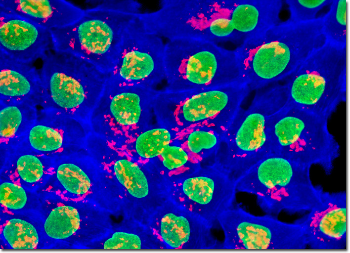
Human Skin Epidermoid Carcinoma Epithelial Cells (A-431)
|
Reducing exposure to ultraviolet light is the best way to avoid developing cutaneous epidermoid carcinoma. Though it sounds simple, carrying out this activity requires constant awareness of one’s environment and taking various preventative measures, such as wearing sunscreen, long sleeve shirts, long pants, and hats when outdoors. Also, shady areas should be chosen whenever possible for outdoor occupations, and special care should be taken of the elderly and young children, who are at greater risk for sunburns due to the delicacy of their skin. However, epidermoid carcinoma is not a disease that only affects the skin. This kind of cancer may develop in most any location of the body where squamous cells are present, including the lungs and other organs. The A-431 cells illustrated above were grown to mid-log phase in adherent monolayer culture, and then fixed, permeabilized, and treated with a cocktail of mouse anti-nuclear pore complex protein NPCP and rabbit anti-giantin (Golgi apparatus) primary antibodies, followed by goat anti-mouse and anti-goat secondary antibodies conjugated to Oregon Green 488 and Alexa Fluor 568, respectively. The filamentous actin cytoskeletal network was simultaneously imaged using Alexa Fluor 350 conjugated to phalloidin. Images were recorded in grayscale with a QImaging Retiga Fast-EXi camera system coupled to an Olympus BX-51 microscope equipped with bandpass emission fluorescence filter optical blocks provided by Omega Optical. During the processing stage, individual image channels were pseudocolored with RGB values corresponding to each of the fluorophore emission spectral profiles. |
© 1995-2025 by Michael W. Davidson and The Florida State University. All Rights Reserved. No images, graphics, software, scripts, or applets may be reproduced or used in any manner without permission from the copyright holders. Use of this website means you agree to all of the Legal Terms and Conditions set forth by the owners.
This website is maintained by our
|