Fluorescence Digital Image Gallery
Embryonic Rat Thoracic Aorta Medial Layer Myoblast Cells (A-10)
|
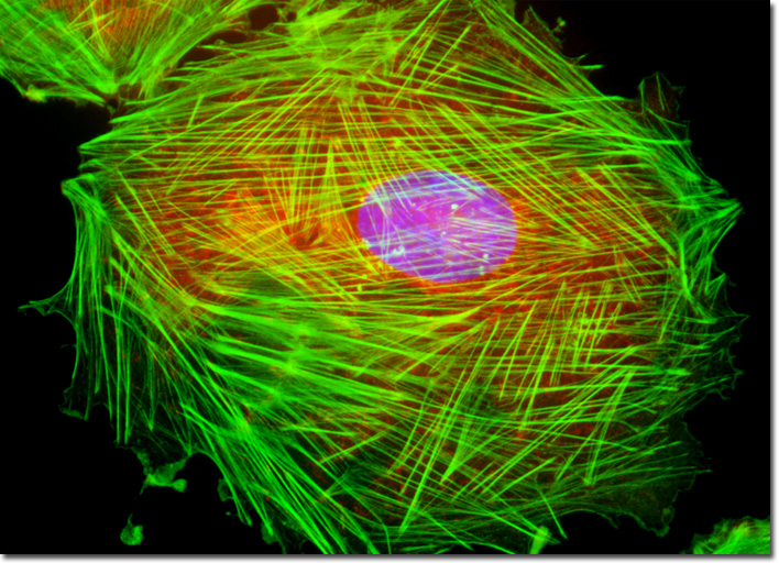
Similar to other blood vessels, the thoracic aorta is comprised of several different layers of tissue, which can generally be observed in a longitudinal section of the arterial wall. The outermost section is the tunica adventitia, which is primarily comprised of collagenous fibers that are arranged in longitudinal spirals, nerves, and blood vessels. This layer also sometimes contains a scattering of ganglion or smooth muscle cells. The middle section, termed the tunica media, contains significant amounts of smooth muscle as well as elastic material that occurs in the form of fenestrated membranes rather than fibers. This medial layer of tissue is the area that was utilized to establish the A-10 cell line. Innermost within the wall of the thoracic aorta is the tunica intima, which is relatively thick, containing connective and smooth muscle tissue as well as an endothelial lining. The culture of rat thoracic aorta (A-10) cells featured in the digital image above was labeled with the enzyme DNase I conjugated to the fluorochrome Texas Red in order to reveal unpolymerized globular (G) actin, which accumulates in the center of the cell. In addition, the specimen was simultaneously stained for DNA with the ultraviolet-absorbing probe DAPI, and for the cytoskeletal filamentous actin network with Alexa Fluor 488 conjugated to phalloidin. Images were recorded in grayscale with a QImaging Retiga Fast-EXi camera system coupled to an Olympus BX-51 microscope equipped with bandpass emission fluorescence filter optical blocks provided by Omega Optical. During the processing stage, individual image channels were pseudocolored with RGB values corresponding to each of the fluorophore emission spectral profiles. |
© 1995-2022 by Michael W. Davidson and The Florida State University. All Rights Reserved. No images, graphics, software, scripts, or applets may be reproduced or used in any manner without permission from the copyright holders. Use of this website means you agree to all of the Legal Terms and Conditions set forth by the owners.
This website is maintained by our
|