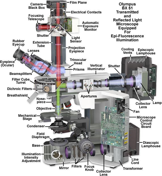
Olympus BX51 Epi-Fluorescence Microscope Cutaway Diagram
|
Microscopes with an upright-style frame are capable of producing fluorescence illumination either through episcopic or diascopic optical pathways, although the latter is rarely used today. Epi-illuminators usually consist of a mercury or xenon lamphouse coupled to a vertical illuminator that is positioned above the main frame in a separate assembly. The microscope nosepiece and transmitted light components (diascopic illuminator, condenser, field diaphragm, filters, etc.) are built into the main frame, while fluorescence components are housed in the vertical illuminator. These illuminators often contain a revolving or sliding turret that houses four to six "cubes" that contain a mixture of interference filters including a barrier filter, dichroic mirror, and an excitation filter. As illustrated above, light emitted from the lamp positioned in the episcopic lamphouse passes through a collector lens and then the field and aperture diaphragms before entering the first interference filter in the cube set, the emission filter. This light is then directed through the objective and onto the specimen by a special dichroic mirror that reflects certain wavelengths while passing others. Secondary fluorescence, emitted by fluorophores residing in the specimen, travels back through the objective and the dichroic mirror before passing through a barrier filter and into the microscope eyepieces or camera system. Contributing Authors William K. Fester and Mortimer Abramowitz - Olympus America, Inc., Two Corporate Center Drive., Melville, New York, 11747. Michael W. Davidson - National High Magnetic Field Laboratory, 1800 East Paul Dirac Dr., The Florida State University, Tallahassee, Florida, 32310. |
© 1995-2022 by Michael W. Davidson and The Florida State University. All Rights Reserved. No images, graphics, software, scripts, or applets may be reproduced or used in any manner without permission from the copyright holders. Use of this website means you agree to all of the Legal Terms and Conditions set forth by the owners.
This website is maintained by our
|