
Differential Interference Contrast Image Gallery
Vorticella Ciliates
There are over 8,000 described ciliates known today. Among them are the bell-shaped protozoans called vorticellas that live in fresh or salt water, often in colonies that may contain between five and several hundred organisms.
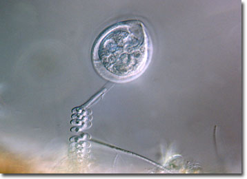
Though they are frequently present in groups, each vorticella possesses its own unciliated stalk. The stalk enables the organisms to attach to aquatic plants and animals, submerged objects, or surface scum. A contractile fiber known as a myoneme is contained within the stalk, causing it to coil up like a spring when it is stimulated or disturbed.
Vorticellas may utilize their cilia to sweep prey into their mouth-like openings. They eat bacteria and other small protozoans, the movement of their cilia creating a water current similar to a vortex, an action that is the basis of their moniker. After food has entered the buccal cavity, the protozoan retracts its contractile stalk and the food is "swallowed."
The primary mode of reproduction for a vorticella is the asexual process of fission. The unicellular creature divides along its length, resulting in two daughter cells. One of the daughter cells retains the original stalk and the other grows temporary cilia along its bottom edge. The organism with additional cilia swims to a new location and eventually loses its extra cilia, grows its own stalk, and affixes to a substrate.