
Differential Interference Contrast Image Gallery
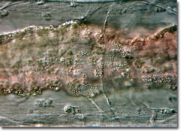
Tubifex Worms
Often referred to as sewage worms, tubifex worms are freshwater annelids that belong to the family Tubificidae. Though they are scientifically described as Tubifex tubifex, their common name resulted from their frequent habitation of polluted, sludge-like waters.
Tubifex worms are capable of thriving in oxygen-poor environments, such as sewage treatment ponds, because they possess a much more efficient manner of assimilating dissolved oxygen than most other organisms. The worms, which generally range in length from 1 to 8.5 centimeters, reside in mud tubes that they create out of a mixture of mud and mucus. However, they often leave their posterior segments outside of the tubes, waving them about and creating a current that enables them to collect any surrounding trace amounts of dissolved oxygen.
As bloodworms, tubifex worms have a relatively high level of hemoglobin and a characteristic bright red color. They are a familiar sight to many aquarium enthusiasts who frequently purchase them as a high-protein food for their favorite fish. The annelids are sold frozen, freeze-dried, or live, though this practice is becoming more rare. Live tubifex worms are not as widely available commercially as they once were due to concerns that they may harbor human pathogens that they acquired in sewage-contaminated waters.