
Differential Interference Contrast Image Gallery
Taenia Tapeworm
Tapeworms belong to the invertebrate class Cestoda and may vary in length from approximately 0.4 inches to 50 feet. Taenia is a genus of tapeworms that are parasites of mammals.
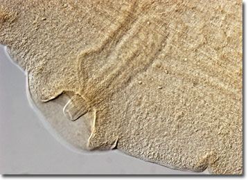
The lifecycle of tapeworms can be complex and may involve multiple hosts, but their anatomy is simple. Species either consist of a single segment or of a succession of identical segments called proglottids and a definite head, known as a scolex. The scolex lacks a mouth, but features suckers and often hooks which are used to attach to the internal organs of a host. Tapeworms also possess a simple nervous system, though they do not have a digestive tract and must absorb nutrients directly from an external layer of tough cuticle. The creatures are typically hermaphrodites and may produce offspring independently.
Tapeworms are capable of infecting a wide variety of animals, including humans, and may cause medical problems if present in large numbers. Taenia solium, for instance, may infest humans if they eat the undercooked pork of an infected pig, and Taenia saginata may be passed on to those who eat contaminated raw beef. Troublesome tapeworm populations can be eliminated from a host, however, with the aid of certain medications. Such treatments generally focus on debilitating the attachment capabilities of the parasites and then washing them away with water and other bodily fluids.